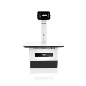
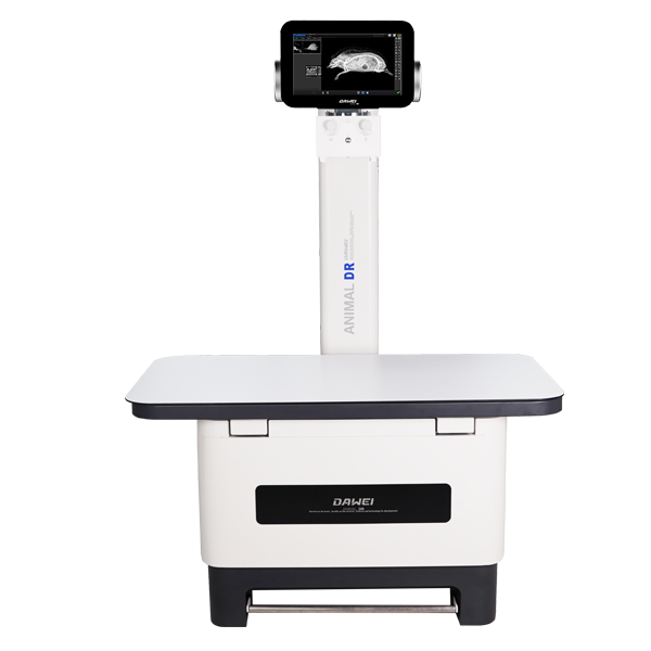
Veterinary Digital Xray Mahine Equipment for Pet Hospital
RV-32A
Applications
Dawei veterinary DR equipment are used for pet digital X-ray examination, which are widely used in all kinds of pet shops due to its excellent production process, low radiation, fast imaging speed, high resolution, safe and stable performance.Whether it's a small exotic pet, a regular dog or cat, or a very large animal, we can meet the requirements of veterinarians to complete all kinds of photography.
1. For musculoskeletal imaging
The 32A animal x-ray machine accurately and rapidly determines skeletal conditions such as fractures, fractures, osteomas, etc.;
2. For chest imaging
The examination of two major organs, lungs and heart, DR images are better than traditional radiography for the display of mediastinum, posterior cardiac area and hidden area under the diaphragm;
3. For abdominal imaging
The 32A animal radiology equipment is used to examine animals for gastrointestinal disorders such as foreign bodies, obstructions, abdominal tumours and reproductive disorders.
4. For X-Ray angiography imaging
It is used in esophagogram and gastrointestinal imaging.
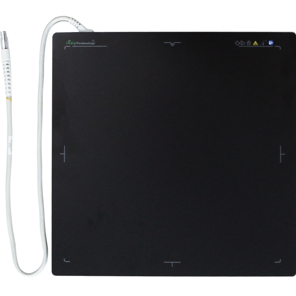
Flat Panel Detector
- Auto Exposure AED Function: The flat panel's unique Auto Exposure Detection function, or AED for short, is sensitive and the flat panel can detect weak exposures.
- Amorphous silicon cesium iodide
- Extremely fast imaging speed
- Large dynamic output range:
- High Stability
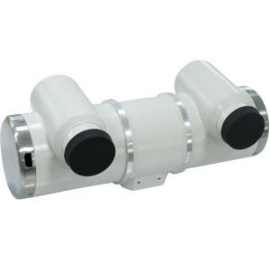
X-Ray Tube Assembly
➣ RV-32A Animal digital X-ray machine equipment:Canon E7239X (imported bulb tube)
- Dual focal spot: small focal spot 1.0mm, large focal spot 2.0mm for the best image resolution and loading power balance
- High thermal capacity: bulb tube thermal capacity up to 140kHU, to achieve more stable continuous exposure performance and better meet the requirements of continuous work.
- Maximum tube current: 340mA for small focus, 570mA for large focus, greatly enhancing the photographic conditions applicable to a variety of animal body types, body position range, to meet the needs of a variety of special photographic loading conditions.
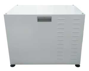
High Voltage Generator
➣ RV-32A veterinary digital X-ray:Canon E7239X (imported bulb tube)
- Dual focal spot: small focal spot 1.0mm, large focal spot 2.0mm for the best image resolution and loading power balance
- High thermal capacity: bulb tube thermal capacity up to 140kHU, to achieve more stable continuous exposure performance and better meet the requirements of continuous work.
- Maximum tube current: 340mA for small focus, 570mA for large focus, greatly enhancing the photographic conditions applicable to a variety of animal body types, body position range, to meet the needs of a variety of special photographic loading conditions.
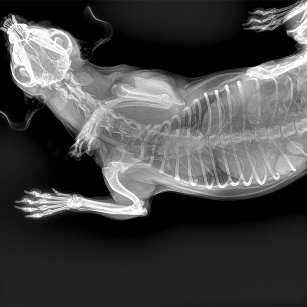
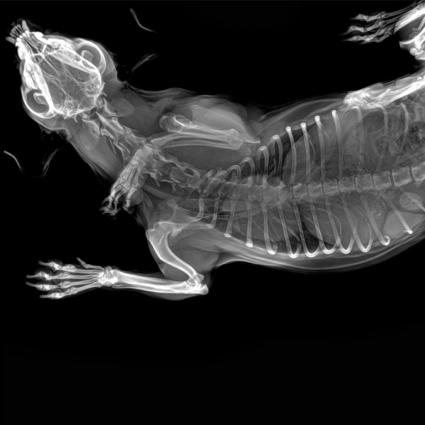
Clinical Images