


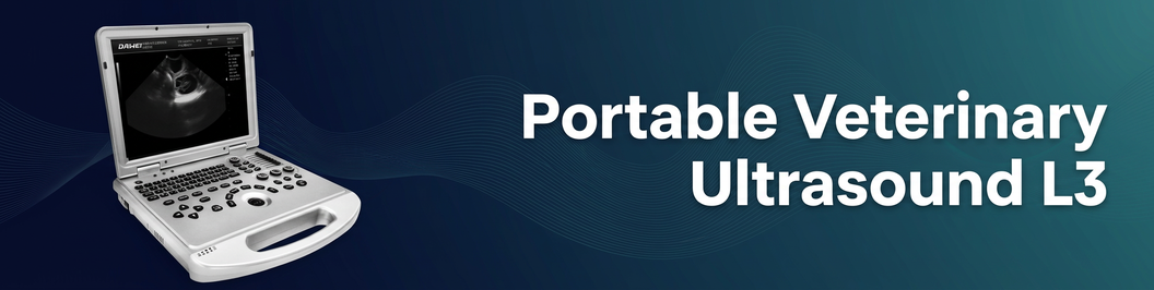
Portable Laptop Animal Reptile Ultrasound Machine Veterinary Ultrasound Scanner
L3-VET
Applications
The L3-VET Veterinary Color Doppler Ultrasound System is a portable laptop-style veterinary ultrasound scanner designed for daily imaging needs in pet hospitals, veterinary clinics, zoos, breeding/reproduction bases, and research institutions.
It combines B-mode, Color Doppler, and Spectral (PW) Doppler with practical image optimization features for fast workflow and confident diagnosis.
Target animals: companion animals and exotics (including reptiles, depending on probe selection and clinical protocol).
For the diagnosis of reptile pregnancy, diagnosis of fish gender.
Key Benefits (Why Clinics Choose L3-VET)
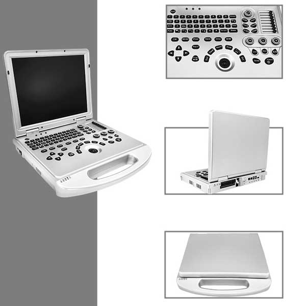
- Laptop-type portability for multi-room clinics, mobile services, and limited spaces
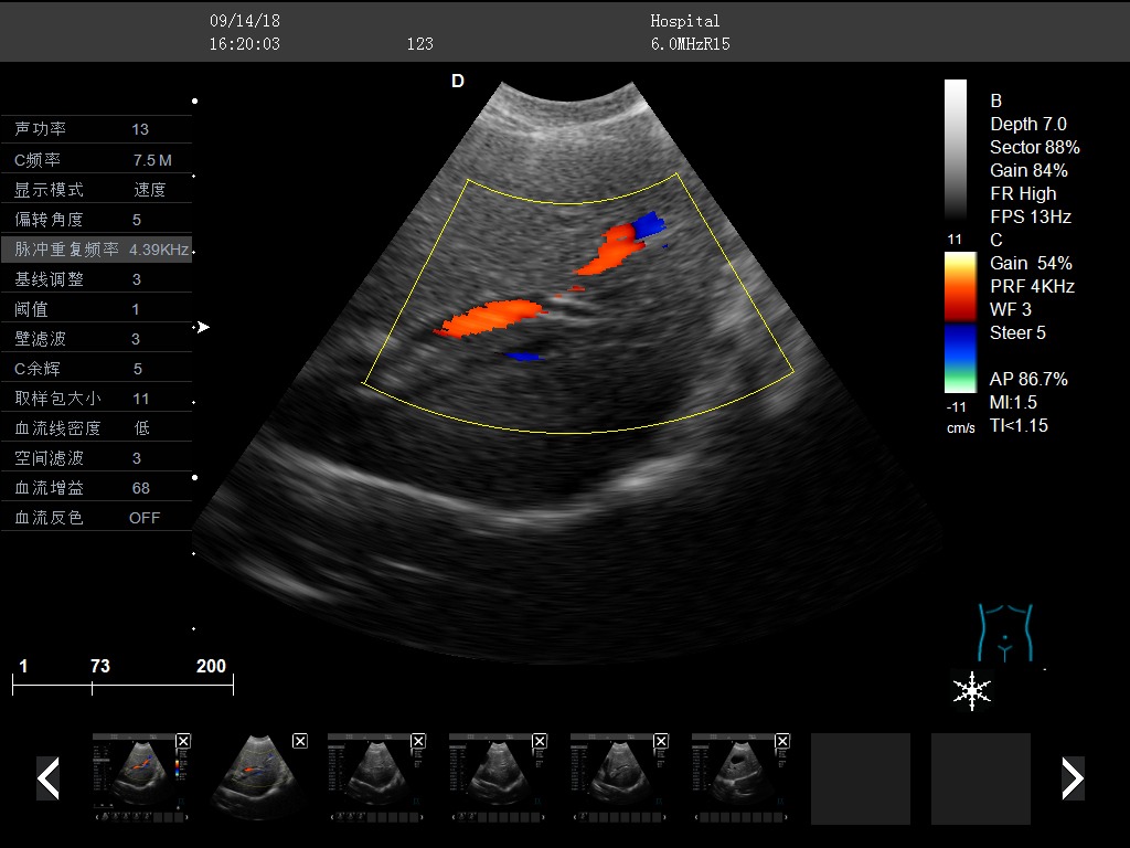
- Color Doppler + PW Doppler to evaluate blood flow and hemodynamics
- Image enhancement tools: THI (Tissue Harmonic Imaging), Compound Imaging, Trapezoidal Imaging
- One-Key Smart Optimization to reduce manual adjustments
- 15-inch monitor for clear viewing during fast-paced exams
- All-in-one clipboard workflow: thumbnails displayed at the bottom for quick save / transfer / delete
- DICOM 3.0 support for PACS connectivity and standardized image management
Imaging Modes & Technologies
Core Modes
- B / C / D real-time three synchronous imaging
- PW (Pulsed Wave) Doppler
- DPDI (Directional Power Doppler Imaging)
- 2B / 4B imaging modes
- B/C split screen (real-time)
Image Quality & Efficiency
- THI (Tissue Harmonic Imaging)
- Compound Imaging
- Trapezoidal Imaging
- One-Key Smart Optimization
- Preset exam conditions for different organs (reduces repeated adjustments)
2D (B-Mode)
| Parameter | Range / Levels |
| Gain | 0–100 (step 1) |
| TGC | 8 segments |
| Dynamic range | 20–280 (20 levels) |
| Pseudo color | 0–11 |
| Sound power | 5%–100% (step 5%) |
| Max focus | ≥ 6 |
| Line density | low / middle / high |
| Noise reduction | 0–14 |
Color Doppler
| Parameter | Range / Levels |
| Color frame correlation | 0–12 |
| Color map | 0–7 |
| Baseline | 11 levels |
| Line density | low / high |
| Filter | 0–5 |
Spectral (PW) Doppler
| Parameter | Range / Levels |
| Angle correction | -80° to 80° |
| Sample volume | 0.5–20 mm |
| Speed scale | 32.8–328 cm/s (varies by probe) |
| Auto calculations | PS, ED, RI, PI, S/D, HR, etc. |
Clinical Applications
Suitable for ultrasound examination needs in:
- Pet hospitals and veterinary clinics
- Zoos and wildlife/exotic animal facilities
- Breeding and reproduction bases
- Universities and scientific research units
Common veterinary use cases include:
- Abdomen (liver, kidney, spleen, bladder, etc.)
- Reproductive evaluation (OB)
- General soft-tissue scanning
- Doppler vascular assessment (as clinically required)

Parameter Control Highlights
Probe
Probe interface ≥ 1
Final probe configuration depends on your quotation and local regulatory requirements.
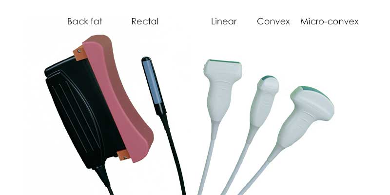
| Probe Type | Detecting Depth | Fundamental Frequency | Harmonic Frequency |
| Convex | 30–255 mm | 2.0 / 2.5 / 3.0 / 3.5 / 4.0 / 5.5 MHz | H4.0 / H5.0 MHz |
| Linear | 20–128 mm | 6.0 / 7.5 / 8.5 / 10.0 / 12.0 MHz | H10.0 MHz |
| Micro-convex (R11) | 30–111 mm | 4.5 / 5.0 / 6.0 / 6.5 / 7.0 / 9.0 MHz | H8.0 MHz |
| Rectal | 20–111 mm | 4.0 / 6.5 / 9.0 MHz | H8.0 MHz |
Quick Specifications
| Item | Specification |
| System type | Veterinary Color Doppler ultrasound diagnostic system |
| Form factor | Laptop type |
| Operating system | Windows 10 |
| Display | 15-inch main monitor |
| Doppler | Color Doppler, DPDI (Directional Power Doppler Imaging), PW (Pulsed Wave Doppler) |
| Imaging sync | B / C / D real-time three synchronous imaging |
| Advanced imaging | THI, Compound Imaging, Trapezoidal Imaging |
| Multi-frame modes | 2B / 4B |
| Presets | Preset exam conditions for different organs |
| Upgrade | On-the-spot upgrade function |
| Probe ports | 1 probe interface |
| Storage | Built-in 128GB hard disk |
| Image formats | BMP / DCM / JPG |
| Movie playback | ≥ 600 frames |
| DICOM | DICOM 3.0, can connect to PACS |
| Interfaces | 4×USB, 1×Audio, 2×RJ-45, 1×HDMI |
| System languages | Chinese / English / French / Russian / Spanish |
Connectivity & Interfaces
| Interface | Quantity |
| USB | 4 |
| Audio | 1 |
| RJ-45 (LAN) | 2 |
| HDMI | 1 |
Measurement, Reporting & Data Management
- General measurements: distance, area, angle, time, slope, HR, speed, acceleration, flow parameters, etc.
- Professional data packages for different organs (examples): Abdomen, OB
- Built-in patient/file management: ID, name, exam number, exam date, search and management
- One-key fast report function
Standard Configuration & Optional Accessories
Standard configuration
- Full digital veterinary color Doppler ultrasound diagnostic system (main unit)
Optional
- Probes: R11 micro-convex / convex / linear / rectal
- Video printer
- Trolley
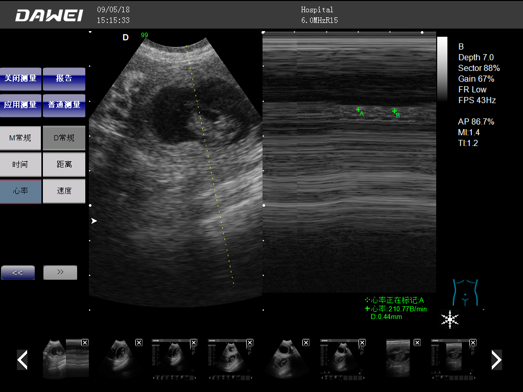
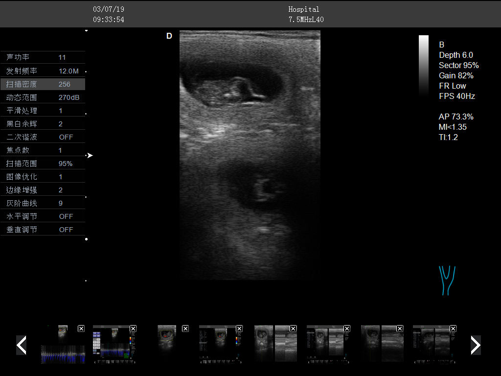
Clinical Images
FAQ
1) What animals can the L3-VET ultrasound scanner be used for?
L3-VET is designed for veterinary imaging in clinics and institutions. With appropriate probe selection and exam presets, it can support small-animal and exotic-animal applications, including reptiles.
2) Does the L3-VET support Doppler?
Yes. It supports Color Doppler, Directional Power Doppler Imaging (DPDI), and Pulsed Wave (PW) Doppler.
3) Can the L3-VET export images to PACS?
Yes. L3-VET supports DICOM 3.0 and can be connected to a PACS system.


