Veterinary Digital X-Ray Machine System | Radiography Medical Imaging Equipment
RV-32B
Applications
The Dawei veterinary x-ray machine is mainly used for the examination of bones, lungs, and foreign bodies of small and medium-sized animals and fish;
- Examination of the skeletal and muscular system
Rapid diagnosis of bone diseases such as fractures, cracks, and osteoma
- Chest examination: Lungs and heart examination
Examination of the lungs and heart
- Abdominal examination
Gastrointestinal foreign bodies, obstruction, abdominal tumors, reproductive system diseases, etc.
32B radiography medical imaging equipment can meet most of the clinical imaging examinations in animal hospitals
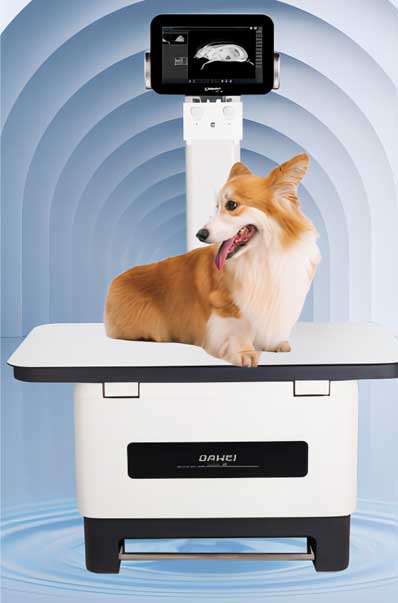
Veterinary digital x-ray machine system & RV-32B Radiography Medical Imaging Equipment
- Flat panel detector: up time 3--5s
- Scintillator: Cesium Iodide
- Pixel matrix: 3072*3072
- Pixel Pitch: 139um
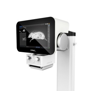
- Display: 15.6 inches
- Screen type: capacitive touch
- High frequency high voltage generator output power: 32KW
- Exposure mA range:10mA-400mA
Performance
Flat Panel Detector
- Comprehensive image production quality has reachedthe forefront level of the domestic industries. It has 139um for pixel size, 3.71p/mm for spatial resolution, 3072*3072 for pixel matrix and millions of pixel imaging
- Large dynamic range (16-bit) and wider grey level can truely satisty the exquisite reproduction of subtle density differences in animal tissue.

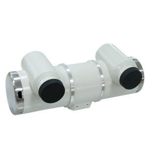
X-Ray Tube Assembly
- Dual focus, high thermal capacity
- Applicable to a wide range of animal body types and positions, to meet the needs of a variety of special photographic loading conditions
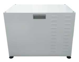
High Voltage Generator
- Output power: 32KW
- Tube voltage range: 40~150kV
- Tube current range: 10mA~400mA (using R'10 or R'20 standard)
- Maximum power: 32KW
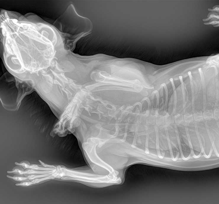
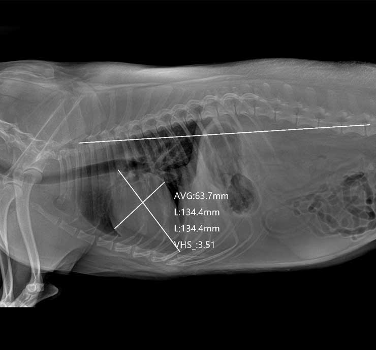
Clinical Images