

Model R Wireless Rectal Veterinary Color Doppler Ultrasound
Model R
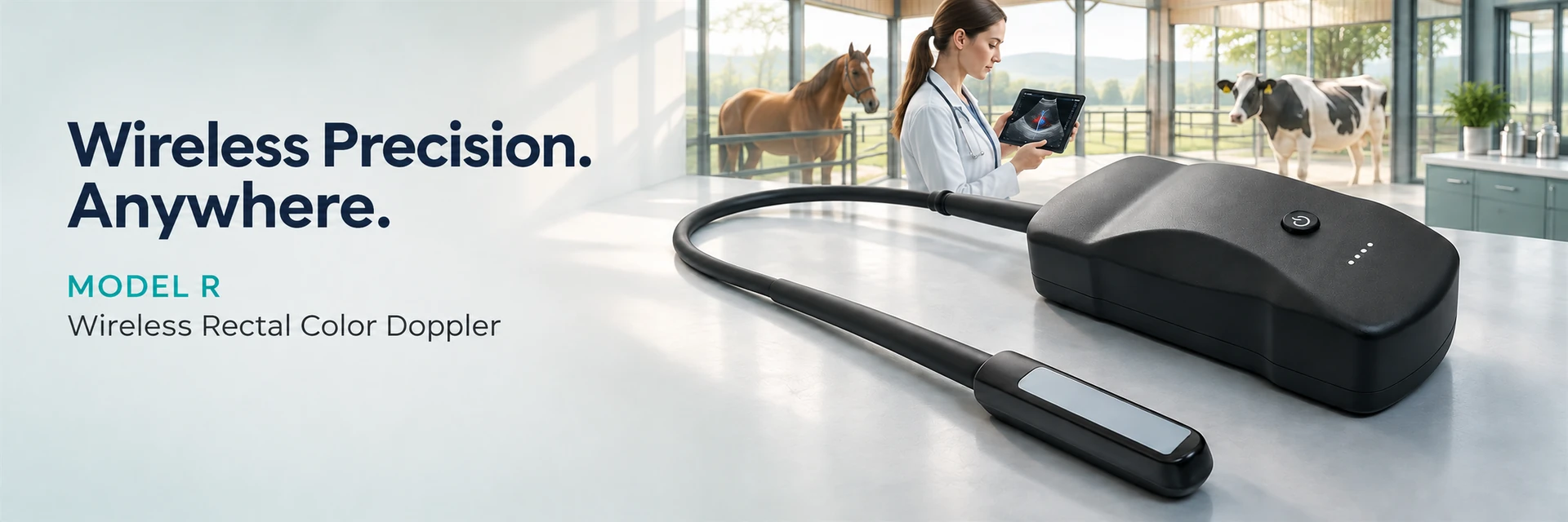
Model R is a wireless-to-display veterinary rectal color Doppler ultrasound designed for cattle and equine reproductive examinations. It combines a 128-element probe, five imaging modes, dual-band Wi-Fi, long B-mode runtime, remote firmware updates, image management, reporting, sharing, and hands-free voice commands on compatible Apple and Samsung devices.
Quick Answers
What is Model R? A handheld veterinary rectal color Doppler ultrasound that sends images wirelessly to a compatible phone, tablet, or M-series Mac.
- Who is it for? Livestock and equine veterinarians, breeding teams, farms, mobile veterinary services, and teaching institutions.
- Which animals are supported? Cattle, horses, sheep, dogs, and pigs.
- Which imaging modes are available?B, B/M, C, PDI, and PW.
- How does it connect? By 2.4 GHz or 5 GHz Wi-Fi, with an open-area range of at least 15 m.
- How long does the battery last?Approximately 9.5 hours in basic B mode; actual runtime varies with use.
- Is it fully cable-free?No. One cable connects the probe to the handheld host; the host sends images wirelessly to the display.
Key Specifications at a Glance
- 128 elements with 64 transmit and 64 receive channels
- B, B/M, C, PDI, and PW imaging modes
- 4.2–12.5 MHz frequency range with 8 selectable steps
- 2.4 GHz / 5 GHz dual-band Wi-Fi
- At least 15 m open-area wireless transmission range
- 10,500 mAh battery; approximately 9.5 hours in basic B mode
- 15 W wireless charging; approximately 7 hours for a full charge
- 189 × 78 × 49 mm; approximately 850 g
- Compatible with iOS, Android, and M-series Mac devices
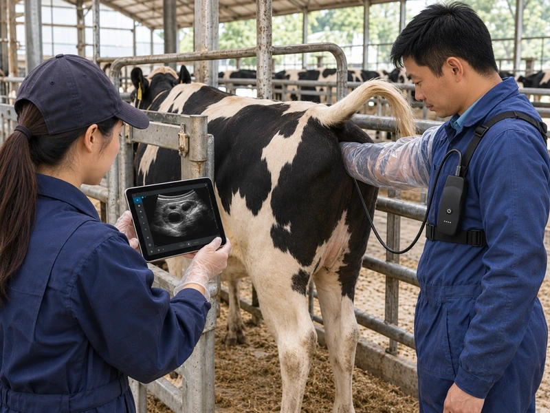
Bovine Reproductive Examination
Designed for cattle pregnancy diagnosis and reproductive assessment. The compact, wearable system allows veterinarians to view ultrasound images on a tablet while maintaining freedom of movement during transrectal examinations.
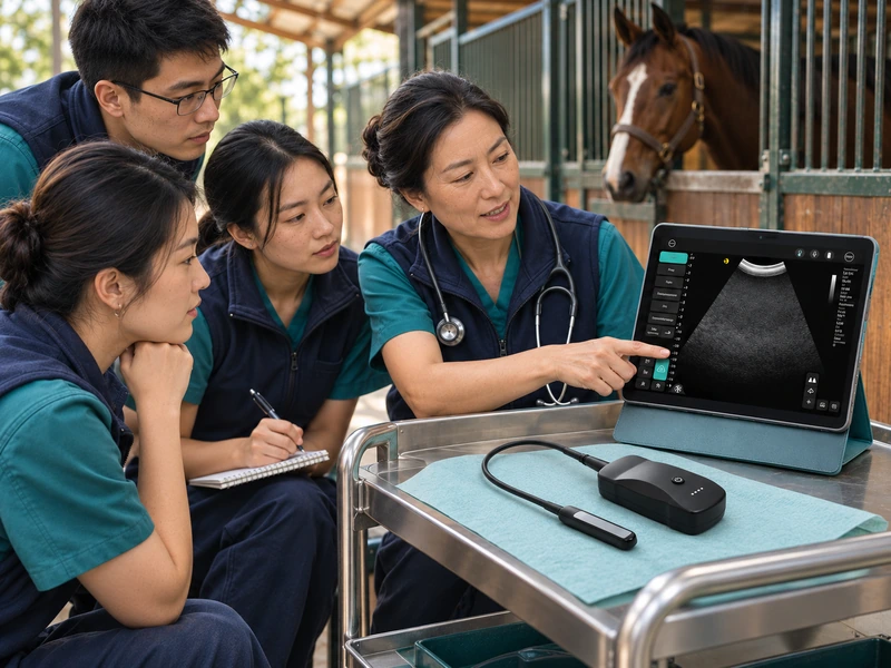
Mobile Practice & Veterinary Training
Ideal for farm visits, equine facilities and veterinary education. Real-time imaging on a tablet makes it easier to review findings, demonstrate scanning techniques and support collaborative clinical decisions.
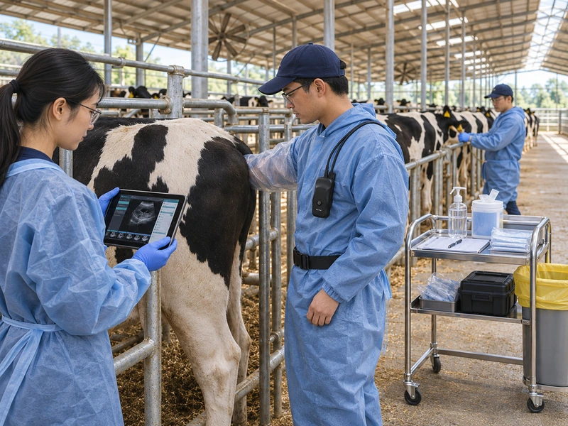
Herd-Side Screening
Suitable for routine reproductive checks on dairy and beef farms. Its portable design supports efficient animal-to-animal examinations without repeatedly moving bulky ultrasound equipment.
Designed for Reproductive Work in the Field
Model R is built for reproductive examinations and mobile workflows where portability, image access, and efficient documentation matter. It can support observation of the reproductive tract, pregnancy assessment, follicle and corpus luteum evaluation, and follow-up imaging in cattle and horses. Clinical interpretation depends on the operator, protocol, and animal condition.
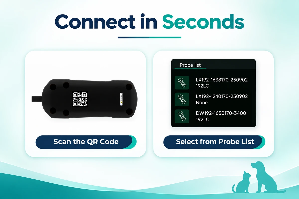
Connect in Seconds
Choose the connection method that fits the situation:
1. Scan the QR code on the device body.
2. Select the device directly from the probe list in the app.
Model R uses 2.4 GHz / 5 GHz dual-band Wi-Fi and supports an open-area range of at least 15 m. The probe remains connected to the handheld host by one cable, while the host connects wirelessly to the display device.
Remote Firmware Upgrade
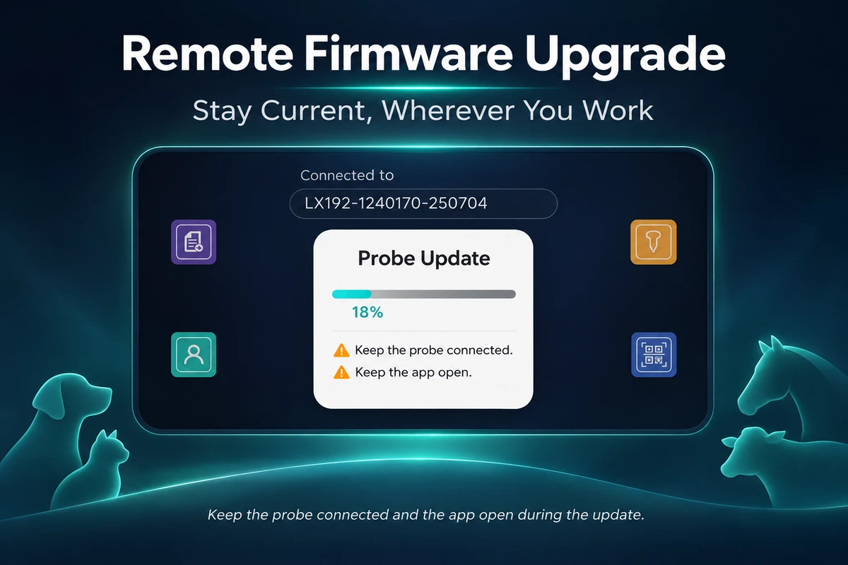
Update the probe firmware through the connected app. During an update, keep the probe connected and leave the app open until the process is complete.
Controls That Adapt to Your Workflow
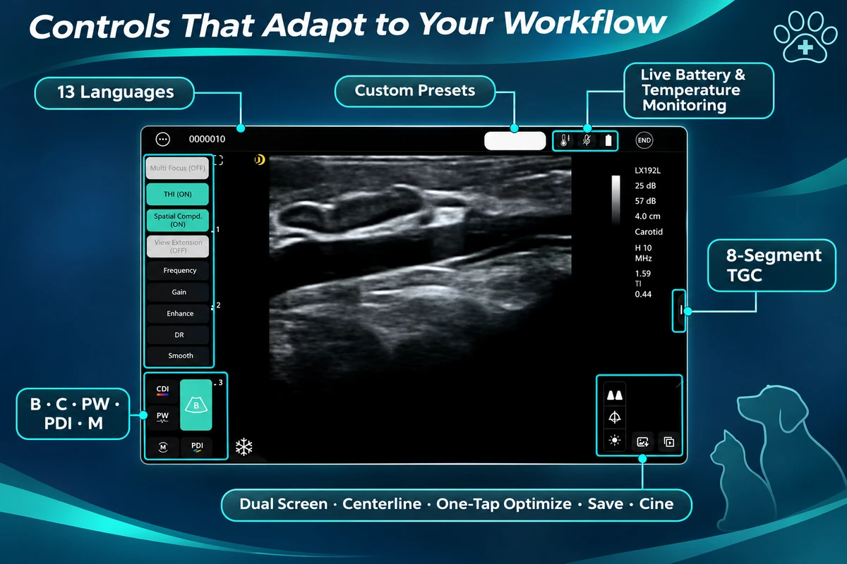
The software interface brings frequently used controls into one workspace:
- B, C, PW, PDI, and M mode selection
- Custom examination presets
- 13 interface languages listed in the supplied presentation
- Live battery and temperature monitoring with warning indicators
- 8-segment TGC with touch adjustment
- Dual-screen view and centerline tools
- One-tap image optimization
- Image saving and cine-loop review
From Scan to Share
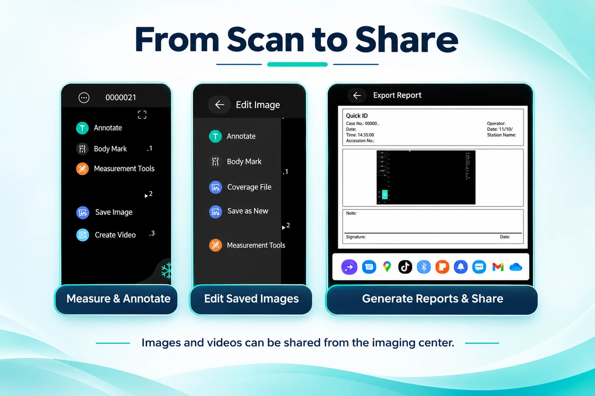
The imaging center supports a connected documentation workflow:
- Add annotations, body marks, and routine measurements
- Reopen and edit saved images
- Save a revised image as a new file
- Generate an examination report
- Print or share images and videos through supported apps
- Export images as JPG, PNG, BMP, or DCM and videos as MP4
- Use DICOM, one-tap sharing, remote meetings, and Zoom integration where available
Hands-Free Voice Control
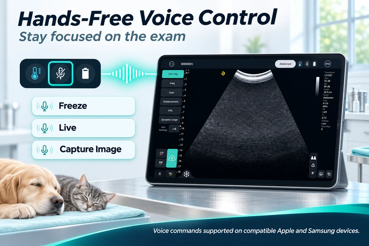
On compatible Apple and Samsung devices, the operator can use three commands:
- Freeze — freeze the live image
- Live — return to live scanning
- Capture Image — save the current image
Compatibility may vary by device model, operating system, app version, and language settings.
Five Imaging Modes for Structural and Blood-Flow Assessment
Model R supports B, B/M, color Doppler (C), power Doppler imaging (PDI), and pulsed-wave Doppler (PW). The 128-element architecture uses 64 transmit and 64 receive channels, with a 4.2–12.5 MHz operating range and an adjustable display depth of 20–188 mm.
Battery Capacity for Mobile Examination Days
The built-in 10,500 mAh battery provides approximately 9.5 hours in basic B mode. The system supports 15 W wireless charging and requires approximately 7 hours for a full charge. Runtime varies with imaging mode, Wi-Fi conditions, display connection, temperature, and battery age.
Compatible Display Devices
Model R supports compatible iOS and Android phones or tablets and Mac computers with M-series chips. Confirm the current supported operating-system versions and device list before ordering.
Typical Applications
Cattle and Equine Reproduction
Reproductive-tract observation, pregnancy assessment, follicle and corpus luteum evaluation, uterine examination, and follow-up imaging.
Herd and Farm Screening
Portable wireless viewing and long B-mode runtime help teams move between animals and examination areas.
Mobile Veterinary Service
Use a compatible phone or tablet as the display to reduce the amount of equipment carried into the field.
Teaching and Remote Collaboration
Save, review, export, and share images or videos for training, case discussion, and remote communication.
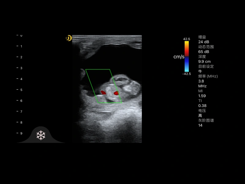
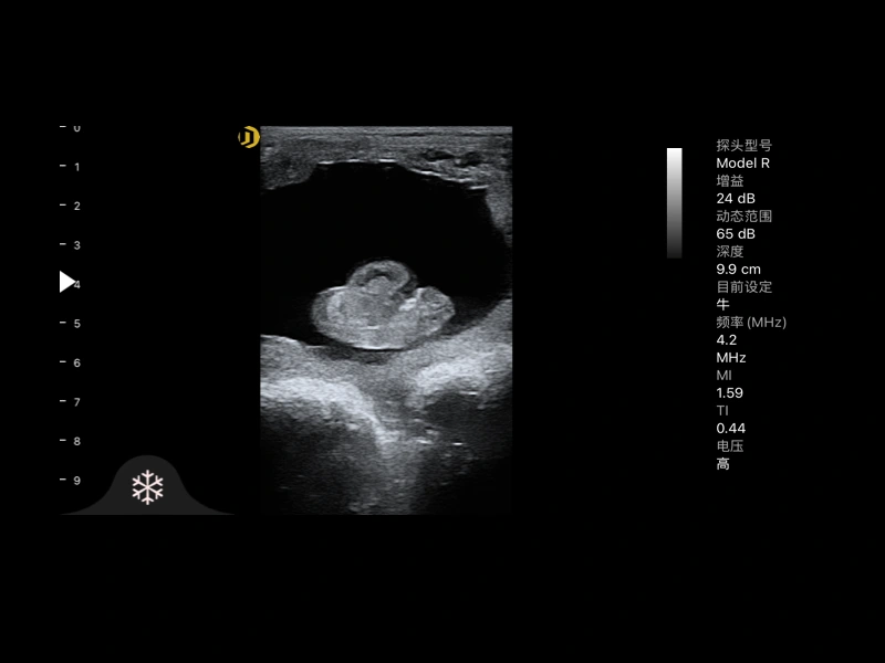
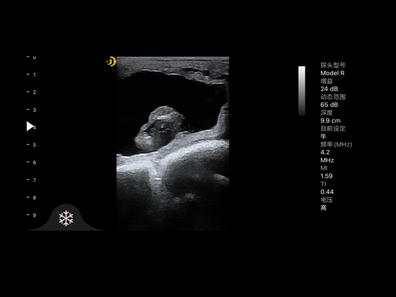
Bovine Pregnancy Ultrasound Imaging with Model R
Frequently Asked Questions
Scan the QR code on the device body or select the device from the probe list in the app.
Model R supports B, B/M, C, PDI, and PW imaging modes.
The technical specification lists iOS, Android, and M-series Mac support. Confirm the current compatibility list before ordering.
Runtime is approximately 9.5 hours in basic B mode. Actual runtime varies with imaging mode, Wi-Fi conditions, display connection, temperature, and battery condition.
Yes. Update the probe firmware through the connected app. Keep the probe connected and the app open during the update.
Yes. The imaging center supports measurements, annotations, body marks, post-editing, report generation, printing, and sharing of supported files.
Compatible Apple and Samsung devices support “Freeze,” “Live,” and “Capture Image.”
Images can be saved as JPG, PNG, BMP, or DCM, and videos as MP4.