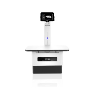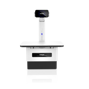


AHD-900 900W Handheld Veterinary DR System
AHD-900
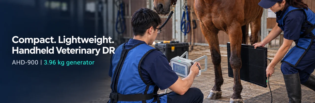
Compact Digital Radiography for Veterinary Care
The AHD-900 combines a 3.96 kg battery-powered X-ray generator with a separate flat panel detector for veterinary digital radiography. With up to 900 W output, a 7-inch touchscreen and an integrated collimator, it brings exposure settings and positioning controls to the source unit.
Built for clinics and veterinary visits, the compact generator can be carried to the examination location and positioned beside the animal. The system serves companion-animal radiography and selected limb examinations in larger animals, with a compatible workstation for image acquisition and review.
What you get from this compact design
- Carry a 3.96 kg generator measuring 221 × 153 × 261 mm between examination locations.
- Select 40–90 kV, 1–10 mA and 0.1–2 s exposure settings on the 7-inch touchscreen.
- Align the examination area using the built-in positioning light and adjustable collimator.
- Capture digital images with the specified 17 × 17-inch-class detector, 139 µm pixel pitch and 16-bit conversion.
A 3.96 kg Generator for Veterinary Visits
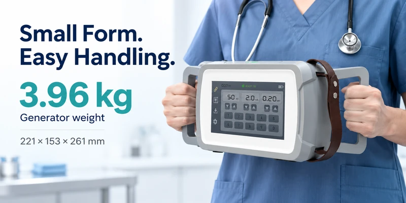
The AHD-900 generator weighs 3.96 kg and measures 221 mm wide, 153 mm high and 261 mm deep. Its compact housing and dual handgrips allow the operator to carry the source to the patient and adjust its position around the examination area.
Battery power at the examination location
The generator operates from a battery supply, so it does not need a mains cable connected during an exposure. This is useful when arranging an examination at a stable, animal-care site or another suitable location away from the clinic. The charger accepts 100–240 V AC input.
Organise the equipment for travel
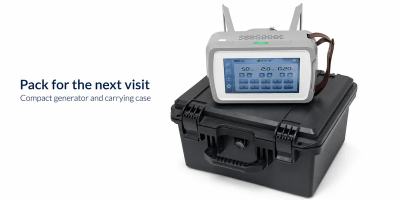
The supplied product package lists a carrying case for transport. Plan the visit around the complete DR setup: generator, detector, acquisition workstation, power supplies and cables. The 3.96 kg specification applies to the generator alone.
For practices with several examination areas, the compact source can also be moved between suitable rooms without moving a full wheeled generator assembly.
Animal Applications and Examination Settings
The AHD-900 is intended for veterinary radiography across companion-animal care and selected larger-animal limb examinations. The table below connects the animal groups in the product presentation with practical examination needs.
| Animal group | Examination focus | Where it fits |
|---|---|---|
| Dogs and cats | Skeletal and limb examinations; chest and abdominal projections suited to patient size. | Companion-animal clinics, selected emergency assessments and follow-up examinations. |
| Rabbits and rodents | Small-animal skeletal and selected body examinations. | Exotic-pet clinics and small-animal practices. |
| Birds and reptiles | Species-appropriate skeletal and selected body projections. | Avian and exotic-animal care. |
| Horses and larger livestock | Selected limb and distal extremity projections. | Equine visits and farm examinations. |
| Wildlife such as deer | Selected limb examinations matched to animal size and access. | Wildlife rescue and veterinary care sites. |
Clinic and emergency examinations
Bring the compact source to the examination position for injury assessment or follow-up radiographs. The separate detector can be arranged behind the target anatomy to suit the required projection.
Surgical checks and postoperative follow-up
The product presentation includes static radiographs for fracture-position checks and postoperative review. These are individual X-ray exposures; this system is not a live fluoroscopy unit.
Mobile visits and animal rescue
A battery-powered generator and carrying case help organise a transportable imaging kit for veterinary visits. Detector and workstation power and connections must also be available at the examination site.
Examination suitability depends on animal size, anatomy, projection and the selected technique. For larger animals, the application focus is selected limbs rather than whole-body coverage.
Exposure Settings and Positioning at the Generator
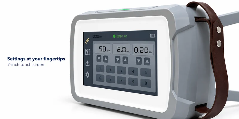
Set exposure parameters on the 7-inch touchscreen
The integrated 800 × 480 touchscreen provides parameter adjustment and preset selection. Operators can call up a stored technique and review the settings at the generator before exposure. This screen controls the source; acquired radiographs are viewed on the workstation.
Adjust the technique to the examination
Select tube voltage from 40 to 90 kV in 1 kV steps, tube current from 1 to 10 mA, and exposure time from 0.1 to 2 seconds. These controls allow the technique to be matched to animal size, anatomy and projection using an appropriate exposure chart. Maximum output is 900 W at 90 kV and 10 mA; setting combinations remain subject to the equipment loading limits.
Align the field before exposing
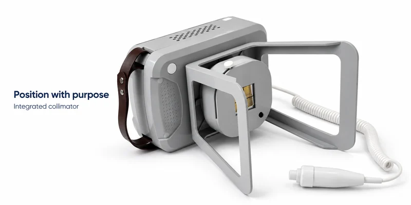
The built-in positioning light shows the intended field before exposure. The adjustable collimator lets the operator narrow or widen that field to match the examination area and detector position. Both functions are integrated into the source housing, so field adjustment takes place where the generator is being positioned.
Trigger exposure with dedicated controls
The product package lists an exposure hand switch and a remote control. These provide exposure-trigger accessories alongside the touchscreen used for parameter settings. Controller operation follows the supplied operating instructions.
Digital Image Acquisition and Flat Panel Detector
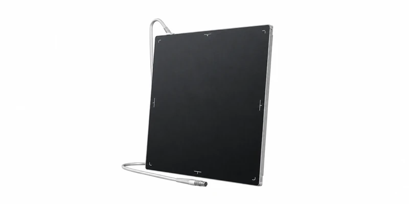
A 17 by 17 inch detector for flexible positioning
The specified detector provides a nominal 430 × 430 mm imaging area. Its square format accommodates different projections and gives room to position the target anatomy. It is a separate component, allowing the source and detector to be arranged on opposite sides of the examination area.
139 micrometre pixels and 16 bit conversion
An amorphous silicon sensor with a cesium iodide scintillator captures the X-ray image. The 3072 × 3072 matrix contains approximately 9.4 million pixels, with a 139 µm pixel pitch and 16-bit A/D conversion. Pixel pitch describes spatial sampling; bit depth describes the available digital intensity levels. Together, these specifications explain how the detector records the image for workstation display and processing.
The generator uses a 40 kHz operating frequency and a 0.8 × 0.8 mm focal spot. The detector has a listed spatial resolution of 3.6 lp/mm. Final detail also depends on positioning, motion, exposure technique and processing.
Image review shortly after exposure
The product presentation lists approximately 3–5 seconds from exposure to image display for its described DR configuration. Prompt image review lets the team check positioning and coverage while preparing the next view. This timing depends on the detector, acquisition software and workstation; confirm it for the quoted configuration.
The 0.1–2 s exposure setting is the X-ray emission time. It is separate from the exposure-to-display interval.
Trigger exposure with dedicated controls
The product package lists an exposure hand switch and a remote control. These provide exposure-trigger accessories alongside the touchscreen used for parameter settings. Controller operation follows the supplied operating instructions.
Clinical Case
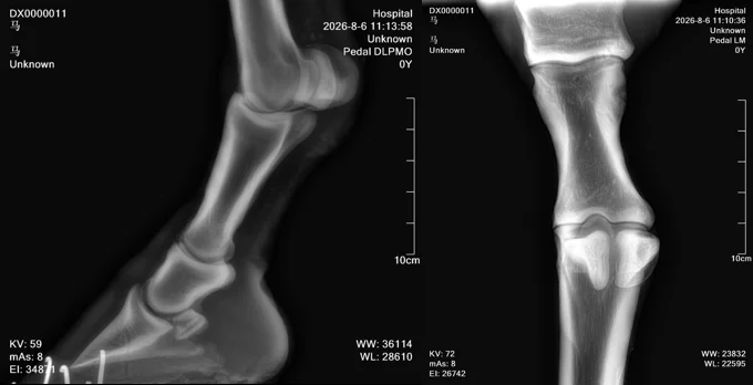
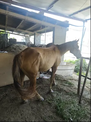
Animal: Horse
Clinical Background: The horse sustained an injury during routine training and required an examination to assess its limb and joint condition.
Examination: Digital radiographic examination of the affected limb and joints using the Dawei Veterinary DR System.
Imaging Findings: The radiographic images provide clear visualization of the bone structures and joint spaces. No obvious abnormalities were reported in this examination.
Clinical Outcome: The examination provided radiographic information to support veterinary assessment of the horse's limb condition.
Application: Equine limb and joint radiography.
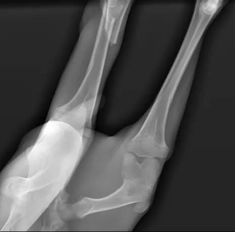
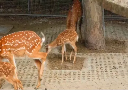
Animal: Sika Deer
Clinical Background: A sika deer sustained an injury to its hind limb after being bitten by another deer. Radiographic examination was required before surgery, during surgical fixation, and for postoperative assessment.
Examination: Digital radiography of the affected hind limb.
Imaging Findings: The X-ray images revealed a femoral fracture.
Diagnosis: Femoral fracture.
Treatment: Digital radiography was used to support fracture assessment and surgical planning, followed by internal fixation surgery.
Clinical Application: Wildlife orthopedic imaging, fracture diagnosis, and surgical assessment.
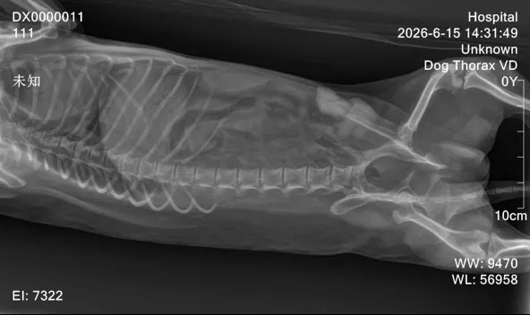

Animal: Dog
Clinical Background: The dog underwent a routine veterinary health examination.
Examination: Digital radiographic examination to assess internal anatomical structures.
Imaging Findings: The radiographic image provides clear visualization of the spine, ribs, pelvis, and surrounding structures.
Clinical Assessment: No obvious abnormalities were reported during the examination.
Treatment / Referral: None reported.
Clinical Application: Routine veterinary radiography and small animal health assessment.
Questions About the AHD 900 Handheld DR System
It describes a veterinary digital radiography setup with a compact handheld X-ray source. For the AHD-900, the generator weighs 3.96 kg; a separate flat panel detector and compatible workstation complete the imaging system.
The application scope includes dogs, cats, rabbits, rodents, birds and reptiles, plus selected limb examinations in horses, livestock and wildlife. The technique and configuration must match the animal size and the required projection.
The generator screen is for exposure settings and preset selection. The acquisition workstation displays the radiographs.
Compatibility depends on the detector model, acquisition software and required DICOM services. Include those details in your enquiry so the proposed configuration can be checked against your existing equipment.
No. Exposure time is selectable from 0.1 to 2 seconds. The presentation describes an approximately 3–5-second exposure-to-display interval for its DR configuration; the quoted detector and workstation determine the applicable performance.
Send your country, animal species, typical animal sizes, required body regions and whether you need a detector, workstation or PACS connection. These details allow the proposed package and price to match your actual examinations.