


L30i Veterinary Ultrasound Machine for Dogs & Cats | Dawei
L30i-VÉT

L30i-VET Veterinary Color Doppler Ultrasound for Small Animals
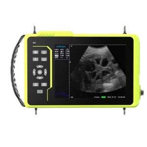
The DAWEI L30i-VET is a laptop-style veterinary ultrasound system developed for pet hospitals, clinics, zoos, breeding facilities and research applications. It combines Color and PW Doppler imaging, veterinary examination presets, flexible probe options and integrated examination data management in a portable platform.
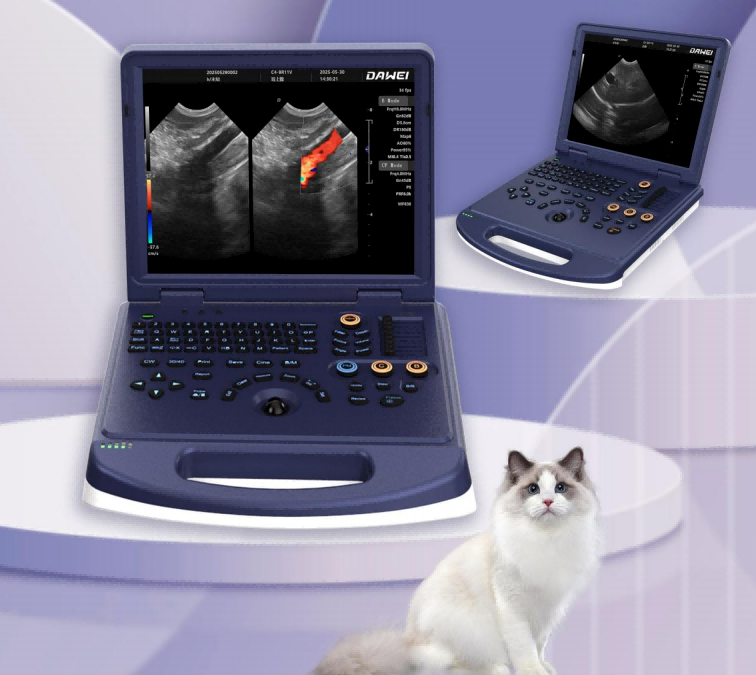
One System for Everyday Small-Animal Ultrasound
The L30i-VET supports a broad range of veterinary ultrasound examinations when configured with the appropriate probe and measurement package.
- Abdominal and urinary: liver, gallbladder, spleen, kidneys, bladder and gastrointestinal structures.
- Reproductive and obstetric: uterus, ovaries, pregnancy monitoring and related measurements.
- Superficial and musculoskeletal: small organs, lymph nodes, mammary tissue, tendons and other superficial structures.
- Vascular and selected cardiac applications: Color and PW Doppler assessment with compatible probes and software packages.
Clear Imaging with Practical Doppler Tools
Imaging functions are organized around routine veterinary examinations, helping operators obtain and review anatomical and blood-flow information efficiently.
Color and Spectral Doppler
Color Doppler, PW spectral Doppler, Directional Power Doppler and real-time triplex imaging support structural and blood-flow assessment.
Advanced 2D Imaging
Spatial compound imaging and Tissue Harmonic Imaging help improve tissue presentation, while trapezoidal imaging expands the field of view for compatible linear probes.
One-Key Optimization
One-key intelligent optimization and preset examination conditions reduce repetitive adjustments during routine scanning.
Flexible Image Controls
Adjustable gain, TGC, dynamic range, focal positions, noise reduction, gray maps and Color/PW parameters support different examination needs.
From Examination to Report
Portable platform: laptop-style structure with a ≥15-inch display and Windows 10 operating system.
Fast review: integrated on-screen clipboard and cine playback of ≥600 frames.
Data storage: ≥128 GB SSD with BMP, JPG and DCM image formats.
System integration: DICOM 3.0 protocol with PACS connectivity.
External interfaces: 6 USB ports, 1 HDMI port and 1 RJ-45 network port.

Choose the Probe for Your Examination Needs
Convex Probe
Eight frequency steps with an imaging depth of 30–255 mm. Suitable for deeper abdominal structures, medium-to-large dogs and obstetric examinations.
Linear Probe
Seven frequency steps with an imaging depth of 20–128 mm. Suitable for superficial structures, small organs, tendons and peripheral vessels.
R11 Microconvex Probe
Five frequency steps with an imaging depth of 30–111 mm. Its compact contact surface is suitable for cats, small dogs and narrow acoustic windows.

Veterinary Measurement Packages
General measurements include distance, area, angle, time, slope, heart rate, velocity, acceleration, RI and PI. Professional packages are available for different organs and examination types.
The system provides at least six report types for abdominal, cardiac, obstetric, small-organ, urinary and vascular examinations.
Standard and Optional Configuration
Standard Configuration
- 1 L30i-VET color Doppler ultrasound main unit
- 1 R11 microconvex probe
Optional Configuration
- Linear probe and other compatible probes
- Video printer
- Ultrasound trolley
L30i-VET Questions
Is the L30i-VET suitable for both dogs and cats?
Yes. The system supports dog, cat and other small-animal examinations. Probe selection should be based on animal size, examination depth and target anatomy.
Which probe is included as standard?
The V3.0C technical configuration lists the R11 microconvex probe as standard. Linear and other compatible probes are optional.
Does the L30i-VET support DICOM?
Yes. It supports the DICOM 3.0 protocol and can connect to a compatible PACS workflow.
Can the system be configured for different clinical applications?
Yes. Available probes, software packages and accessories can be selected according to the animals and examinations performed by the clinic.
Request an L30i-VET Configuration
Tell us the animal species, typical body size and examinations performed by your clinic. DAWEI can recommend an appropriate probe and system configuration.
Product functions and available options may vary by configuration. Please confirm the latest specifications and supplied accessories before ordering.
L30i-VET Veterinary Color Doppler Ultrasound
A laptop-style veterinary ultrasound platform for pet hospitals, clinics, zoos, breeding facilities and research applications. The system combines Color and PW Doppler imaging, veterinary measurement packages and integrated examination data management.
| Specification | L30i-VET |
|---|---|
| System and workflow | |
| Product type | Veterinary color Doppler ultrasound diagnostic system |
| System design | Portable laptop-style structure |
| Recommended environments | Pet hospitals, veterinary clinics, zoos, breeding and reproduction facilities, and research institutions |
| Operating system | Windows 10 |
| Display | ≥15-inch monitor |
| System languages | Chinese, English, French, Russian and Spanish |
| Probe connectors | ≥1 probe connector |
| Workflow functions | Integrated on-screen clipboard, examination presets, one-key intelligent optimization and on-site system upgrade support |
| Imaging and Doppler | |
| Imaging modes | B, 2B, 4B, Color Doppler, PW spectral Doppler, Directional Power Doppler and real-time triplex imaging |
| Advanced imaging | Spatial compound imaging, Tissue Harmonic Imaging (THI) and trapezoidal imaging |
| 2D image controls | Gain 0–100; 8-segment TGC; dynamic range 20–280 dB in 14 visible levels; pseudocolor 0–11; acoustic power 5%–100% in 5% steps |
| 2D processing | Gray map 0–7; filter 0–4; scan range 50%–100%; frame correlation 0–4; noise reduction 0–14; high/medium/low scan-line density |
| Focusing and body marks | Up to 6 fully movable focal points and ≥18 body-position markers |
| Color controls | Color frame correlation 0–12; color maps 0–7; color inversion; 11-level baseline; high/low line density; wall filter 0–5; synchronized B/C split-screen display |
| PW Doppler controls | Angle correction -80° to +80°; sample volume 0.5–20 mm; 11-level baseline; velocity scale 32.8–328 cm/s; ≥4 display layouts |
| Spectral analysis | Real-time automatic and manual spectral tracing with automatic calculation of PS, ED, PI, RI, S/D and HR |
| Spectral processing | Spectrum pseudocolor 0–5; gray map 0–7; wall filter 0–8; dynamic range 10–90 dB in steps of 5; noise reduction 0–60; volume 0–100 |
| Probe options | |
| Convex probe | 2.0, 2.5, 3.0, 3.5, 4.0 and 5.5 MHz; harmonic frequencies H4.0 and H5.0 MHz; 8 frequency steps; imaging depth 30–255 mm |
| Linear probe | 6.0, 7.5, 8.5, 10.0 and 12.0 MHz; harmonic frequencies H9.0 and H10.0 MHz; 7 frequency steps; imaging depth 20–128 mm |
| R11 microconvex probe | 4.0, 6.0, 7.0 and 8.0 MHz; harmonic frequency H8.0 MHz; 5 frequency steps; imaging depth 30–111 mm |
| Measurements and veterinary packages | |
| General measurements | Distance, area, angle, time, slope, heart rate, velocity, acceleration, Resistance Index (RI) and Pulsatility Index (PI) |
| Veterinary measurement packages | Obstetric, hepatobiliary, renal, bladder, cardiac, gastrointestinal, uterine, adrenal, pancreatic and vascular applications |
| Measurement customization | Adjustable measurement-line color and type, including active and completed colors; adjustable result position and font size |
| Data management and connectivity | |
| Internal storage | ≥128 GB SSD |
| Cine playback | ≥600 frames |
| Image formats | BMP, DCM and JPG |
| Patient archive | Stores and searches examination data by ID, pet name, pet number and owner name |
| Report templates | ≥6 report types: abdominal, cardiac, obstetric, small-organ, urinary and vascular ultrasound reports |
| DICOM and PACS | DICOM 3.0 protocol with PACS connectivity |
| Interfaces | 6 USB ports, 1 HDMI port and 1 RJ-45 network port |
| Configuration | |
| Standard configuration | 1 L30i-VET color Doppler ultrasound main unit and 1 R11 microconvex probe |
| Optional configuration | Linear probe and other compatible probes, video printer and ultrasound trolley |
Specifications and available options may vary by configuration. Please contact DAWEI to confirm the latest configuration before ordering.
Related Products
-
50 kW Large Animal Veterinary X-ray System RV-50A
-

Cattle Equine Ovum Pick-UP(OPU) Ultrasound System
-
Basic Laptop Full Digital Veterinary Ultrasound...
-
Wireless Ultrasound Color Doppler Device Probe ...
-

Livestock Ultrasound Machine for Cattle, Horse,...
-
Waterproofing Handheld Cattle Equine Portable V...


