


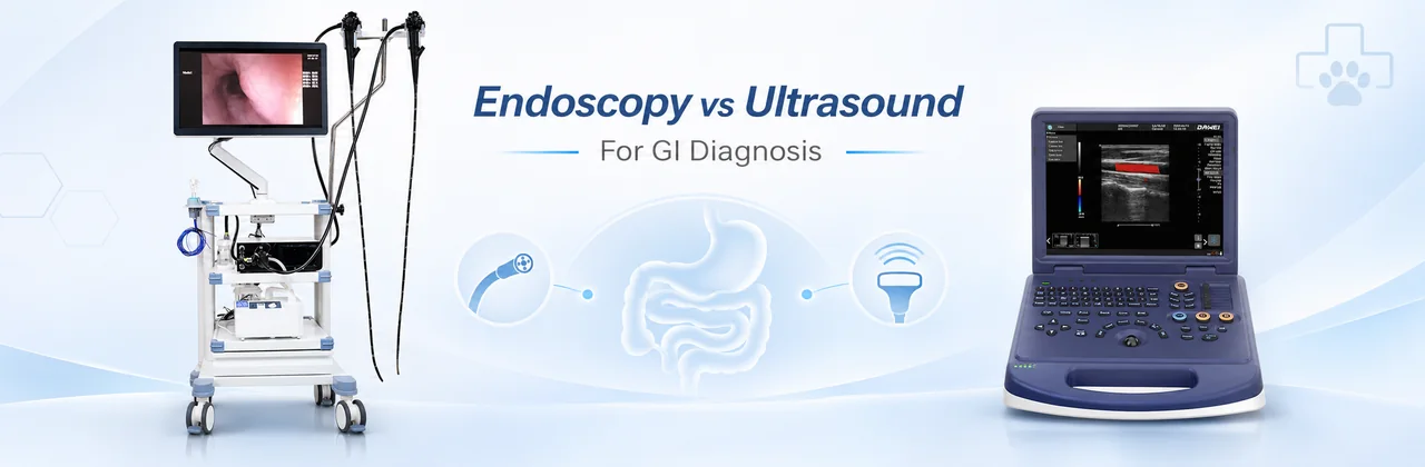
When a dog or cat experiences persistent vomiting, diarrhea, unexplained weight loss, or suspected gastrointestinal disease, veterinarians may use different diagnostic tools to investigate the underlying cause.
Two important methods are veterinary abdominal ultrasound and gastrointestinal endoscopy.
Although both are used to evaluate digestive disorders, they provide different types of clinical information.
Ultrasound is useful for assessing gastrointestinal wall thickness, wall layering, intestinal movement, and surrounding abdominal organs. Endoscopy allows direct examination of the gastrointestinal mucosal surface and, when indicated, collection of tissue samples for histopathology.
So, which method is better?
The answer depends on the animal’s clinical signs, suspected disease, and diagnostic objectives. In many cases, ultrasound and endoscopy complement each other rather than compete.
This guide explains their differences, clinical applications, limitations, and how veterinarians can select suitable equipment for small-animal gastrointestinal examinations.
1. What Is Veterinary Gastrointestinal Ultrasound?
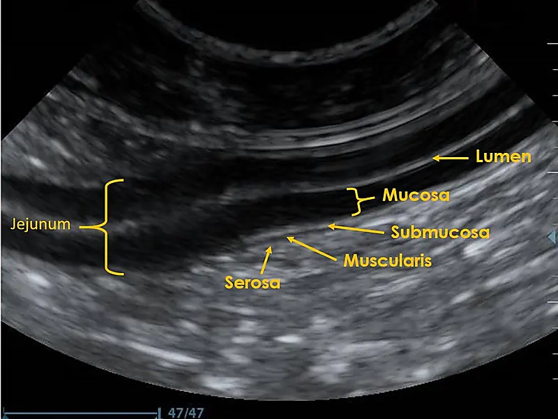
Veterinary gastrointestinal ultrasound is a noninvasive imaging technique that uses high-frequency sound waves to evaluate the digestive tract and surrounding abdominal structures.
During the examination, a veterinarian places an ultrasound transducer against the animal’s abdomen. The transducer sends and receives sound waves, which are converted into real-time images.
Unlike endoscopy, ultrasound evaluates structures from outside the gastrointestinal tract.
What Can Ultrasound Detect in Dogs and Cats?
Abdominal ultrasound can provide information about:
- Gastrointestinal wall thickness: Identifying areas of abnormal thickening or thinning.
- Intestinal wall layering: Evaluating whether the normal layered appearance is preserved or altered.
- Intestinal motility: Observing movement of accessible intestinal segments.
- Suspected intestinal obstruction: Identifying bowel dilation, abnormal contents, or associated changes.
- Intussusception: Detecting characteristic abnormalities when one segment of intestine telescopes into another.
- Abdominal masses: Evaluating certain masses and their relationship to nearby structures.
- Free abdominal fluid: Identifying abnormal fluid accumulation.
- Surrounding organs: Examining the liver, pancreas, spleen, kidneys, lymph nodes, and other abdominal structures.
Ultrasound is particularly valuable when clinical signs suggest that the problem may involve more than the gastrointestinal mucosal surface.
However, gastrointestinal gas, patient body condition, and operator experience can affect image quality.
A normal ultrasound examination also does not exclude every gastrointestinal disease, particularly microscopic mucosal disorders.
Veterinary Ultrasound Equipment for GI Examinations
A portable color Doppler ultrasound system, such as the Dawei L30i-VET pet ultrasound machine, can support abdominal examinations in dogs and cats when equipped with appropriate probes.
The L30i-VET provides B-mode imaging, color Doppler functions, and compatible probe options for different small-animal examination requirements.
For gastrointestinal assessment, the choice between microconvex and high-frequency linear probes depends on the patient’s size, target depth, and required image detail.
Veterinarians should select the transducer and imaging parameters according to the clinical examination rather than relying on one preset for every patient.
2. What Is Veterinary Gastrointestinal Endoscopy?
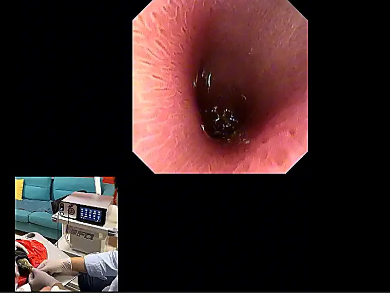
Veterinary gastrointestinal endoscopy is a diagnostic technique that uses a flexible endoscope equipped with a camera and illumination system to examine the internal surface of accessible digestive organs.
The endoscope is introduced through a natural body opening, allowing the veterinarian to inspect gastrointestinal mucosa directly.
Depending on the procedure, gastrointestinal endoscopy may include:
- Esophagoscopy: Examination of the esophagus
- Gastroscopy: Examination of the stomach
- Duodenoscopy: Examination of the duodenum
- Colonoscopy: Examination of the colon
- Ileoscopy: Examination of accessible portions of the ileum
Endoscopy is especially valuable when the clinical question involves mucosal disease, targeted biopsy, or a foreign body that can potentially be retrieved endoscopically.
What Can Veterinary Endoscopy Detect?
Gastrointestinal endoscopy can help veterinarians identify:
Gastric and intestinal mucosal lesions
Endoscopic imaging may reveal visible inflammation, erosions, ulceration, bleeding, or other mucosal abnormalities.
Esophageal and gastric foreign bodies
In suitable cases, endoscopy allows visualization and removal of accessible foreign objects using compatible retrieval instruments.
Abnormal mucosal growths
Endoscopy can identify visible polyps, masses, and other changes affecting the gastrointestinal surface.
Conditions requiring tissue biopsy
When clinically indicated, endoscopic biopsy provides tissue samples for histopathological examination.
This can be important when investigating selected chronic gastrointestinal diseases.
However, not every animal with vomiting or diarrhea requires an endoscopic biopsy. The decision should follow clinical assessment, laboratory testing, imaging, and appropriate initial treatment or dietary trials when indicated.
Veterinary Endoscopy Equipment for Dogs and Cats
The Dawei AEG-200 veterinary endoscopy system is designed for gastrointestinal examinations in small and large animals.
Its published configurations include:
- 6.0 mm and 8.8 mm flexible scope options
- 1000 mm and 1500 mm working lengths
- 140° field of view
- HD imaging
- Integrated light source and image-management functions
These options allow veterinary clinics to consider different patient sizes and examination requirements.
The selected endoscope, working channel, and compatible accessories must be appropriate for the intended diagnostic or therapeutic procedure.
For smaller anatomical passages, an ultrathin endoscope may be considered, although its working-channel dimensions can limit accessory selection and therapeutic capabilities.
3. Veterinary Endoscopy vs Ultrasound: Key Differences
Both techniques are important in veterinary gastrointestinal diagnosis, but their capabilities are different.
| Feature | Abdominal Ultrasound | Gastrointestinal Endoscopy |
|---|---|---|
| Examination method | Imaging through the abdominal wall | Direct visualization through a flexible scope |
| Main diagnostic strength | Abdominal anatomy and gastrointestinal wall assessment | Mucosal visualization and targeted biopsy |
| GI wall thickness | Can be assessed | Not reliably measured through routine visual endoscopy |
| Intestinal wall layering | Can be evaluated | Cannot directly assess full wall layering |
| Mucosal surface | Cannot be directly visualized | Can be directly examined |
| Surrounding abdominal organs | Can be assessed | Cannot directly assess surrounding organs |
| Tissue sampling | Image-guided sampling may be possible with additional procedures | Mucosal biopsy through compatible instruments |
| Foreign body assessment | Can help identify certain foreign bodies and obstruction | Can inspect and sometimes retrieve accessible foreign bodies |
| Anesthesia | Often possible without general anesthesia | Usually requires general anesthesia in dogs and cats |
| Main limitations | Gas, body condition, and operator dependence | Limited reach and limited assessment beyond the mucosa |
Key takeaway: Ultrasound provides information about the gastrointestinal wall and surrounding abdomen, while endoscopy provides a direct view of the internal mucosal surface.
Neither method can replace the other in every clinical situation.
4. When Is Ultrasound More Useful for Gastrointestinal Diagnosis?
Abdominal ultrasound is often a valuable examination when a veterinarian needs to investigate the structure of the digestive tract or evaluate other abdominal organs.
Suspected Intestinal Obstruction
A dog with vomiting, abdominal discomfort, and reduced appetite may have a gastrointestinal obstruction.
Ultrasound can help assess intestinal dilation, abnormal contents, wall changes, and other findings associated with obstruction.
Abdominal radiography is also important in many suspected obstruction cases, and the most appropriate imaging sequence depends on clinical urgency.
Gastrointestinal Wall Abnormalities
Ultrasound can identify changes in intestinal wall thickness and layering.
These findings may raise concern for inflammatory, infiltrative, or neoplastic disease.
However, ultrasound alone generally cannot establish the microscopic cause of these changes.
Further investigation, including tissue sampling when appropriate, may be necessary.
Abdominal Masses and Surrounding Organs
A gastrointestinal complaint may originate from structures outside the intestinal lumen.
Ultrasound allows veterinarians to assess accessible surrounding organs, including the pancreas, liver, spleen, and abdominal lymph nodes.
This broader examination is one of the main advantages of abdominal ultrasonography.
Intestinal Motility and Fluid
Real-time imaging allows observation of intestinal movement and detection of abdominal fluid.
These findings can contribute to clinical assessment but must be interpreted alongside patient history, physical examination, and other test results.
5. When Is Endoscopy More Useful?
Endoscopy is particularly valuable when the veterinarian needs to observe mucosal abnormalities directly, collect targeted biopsies, or perform selected minimally invasive interventions.
Persistent Gastrointestinal Signs
Dogs and cats with persistent vomiting, diarrhea, or weight loss may require additional investigation when initial testing and appropriate treatment do not establish the cause.
Endoscopy may be recommended when mucosal disease is suspected and tissue biopsy is necessary.
The timing of endoscopy depends on the patient’s condition, diagnostic findings, and response to previous management.
Suspected Gastric Ulcers
Gastrointestinal ultrasound may identify some associated abnormalities, but it is less sensitive for many nonperforated mucosal ulcers.
Endoscopy enables direct visualization of accessible mucosal ulceration and associated lesions.
Foreign Body Retrieval
Endoscopy can sometimes remove foreign objects from the esophagus or stomach without open abdominal surgery.
However, it is not appropriate for every foreign body.
Objects located farther along the intestine, suspected perforation, complicated linear foreign bodies, or severe obstruction may require surgery.
Gastrointestinal Biopsy
Endoscopic biopsy allows veterinarians to collect mucosal tissue for histopathological evaluation.
This can support the investigation of selected inflammatory or neoplastic gastrointestinal conditions.
A limitation is that routine endoscopic biopsy usually samples superficial mucosal tissue.
When disease involves deeper intestinal layers or inaccessible sites, other sampling methods may be required.
6. Which Examination Should Be Considered for Common Clinical Problems?
The selection of diagnostic tools should begin with the suspected condition rather than with equipment availability alone.
| Clinical Situation | Ultrasound | Endoscopy |
|---|---|---|
| Unexplained vomiting | Useful for abdominal and structural assessment | Consider when accessible mucosal disease or a retrievable foreign body is suspected |
| Chronic diarrhea | May reveal bowel wall or associated abdominal changes | Consider for mucosal biopsy when indicated after clinical workup |
| Suspected gastric ulcer | Can reveal some associated abnormalities | Direct mucosal examination is especially useful |
| Suspected gastric foreign body | Can contribute to imaging assessment | May allow retrieval if the object is safely accessible |
| Suspected intestinal obstruction | Useful alongside radiography | Limited role; surgery may be required |
| Suspected intestinal mass | Evaluates wall and surrounding structures | May permit mucosal biopsy if the lesion is accessible |
| Unexplained abdominal fluid | Useful for detection and evaluation | Generally not the primary method |
Clinical decisions should be individualized. These examples illustrate common diagnostic roles and are not a fixed examination sequence for every animal.
7. Can Ultrasound and Endoscopy Be Used Together?
Yes. In many gastrointestinal cases, abdominal ultrasound and endoscopy provide complementary clinical information.
For example, consider a dog with persistent vomiting and unexplained weight loss.
A veterinarian may first perform a physical examination, laboratory testing, and abdominal imaging.
Ultrasound could help evaluate intestinal wall structure, abdominal lymph nodes, the pancreas, and other accessible organs.
If the findings or clinical history suggest a condition requiring mucosal evaluation, endoscopy may then be considered.
Endoscopy can provide direct visualization and targeted biopsy samples from accessible gastrointestinal segments.
An Illustrative Diagnostic Workflow
Step 1: Clinical assessment
Review patient history, clinical signs, physical examination findings, and appropriate laboratory tests.
Step 2: Abdominal imaging
Use radiography and/or ultrasound as indicated to assess abdominal anatomy, suspected obstruction, or structural abnormalities.
Step 3: Determine whether endoscopy is indicated
Consider endoscopy when direct mucosal examination, biopsy, or removal of an accessible foreign body would influence diagnosis or treatment.
Step 4: Integrate the findings
Interpret imaging, endoscopic observations, and histopathological results together with the patient’s clinical condition.
This is an illustrative workflow, not a universal protocol. Some emergencies require immediate intervention, while some chronic gastrointestinal cases may benefit from dietary or medical management before endoscopy.
8. Choosing Veterinary Imaging Equipment for Gastrointestinal Examinations
For veterinary hospitals and clinics, selecting suitable gastrointestinal diagnostic equipment depends on patient size, expected case volume, and clinical services.
What to Consider When Choosing an Ultrasound System
For canine and feline abdominal examinations, consider:
- Image quality and gastrointestinal wall detail
- Appropriate microconvex and linear probe options
- Adjustable depth, gain, and focal settings
- Real-time image recording and review
- Suitable equipment size for the clinic
The Dawei L30i-VET is a portable color Doppler ultrasound platform designed for small-animal examinations, including abdominal imaging.
What to Consider When Choosing an Endoscopy System
For gastrointestinal endoscopy, important considerations include:
- Scope diameter for different patient sizes
- Working length for the intended anatomical reach
- Working-channel diameter and accessory compatibility
- Image quality and illumination
- Air, water, and suction functions
- Cleaning, disinfection, and storage requirements
- Operator training and anesthesia support
For routine gastrointestinal visualization in dogs and appropriately selected patients, the Dawei AEG-200 provides flexible scope configurations.
The Dawei AEG-180 portable veterinary endoscope is another option for selected small-animal and narrow-anatomy applications, with ultrathin scope configurations. Its smaller working channel may limit certain biopsy or retrieval procedures.
Clinics comparing endoscopy configurations can also review our guide to choosing endoscope equipment for veterinary surgery.
9. Frequently Asked Questions
Is ultrasound or endoscopy better for a vomiting dog?
It depends on the suspected cause. Ultrasound is useful for evaluating abdominal structures, intestinal wall changes, and certain obstructions. Endoscopy may be more useful when direct inspection of the upper gastrointestinal mucosa, biopsy, or retrieval of an accessible foreign body is needed.
Can ultrasound detect gastrointestinal foreign bodies in dogs?
Ultrasound can identify some foreign bodies and associated obstruction findings. However, not all objects are visible, and abdominal radiography may also be necessary. A negative ultrasound does not completely exclude a foreign body.
Can endoscopy replace abdominal ultrasound?
No. Endoscopy primarily examines accessible mucosal surfaces, while ultrasound evaluates gastrointestinal wall structures and surrounding abdominal organs. They provide different information.
Can abdominal ultrasound diagnose inflammatory bowel disease in dogs?
Ultrasound may reveal findings associated with chronic intestinal disease, but it cannot independently establish a definitive diagnosis of canine chronic inflammatory enteropathy or distinguish every inflammatory condition from neoplasia. Diagnosis requires interpretation of the broader clinical workup, and biopsy is appropriate in selected cases.
Does veterinary endoscopy require general anesthesia?
Gastrointestinal endoscopy in dogs and cats generally requires general anesthesia for patient safety, airway protection, and controlled examination. The veterinarian should assess anesthetic risks before the procedure.
Can endoscopy remove intestinal foreign bodies?
Endoscopic retrieval is mainly useful for selected accessible objects, particularly in the esophagus and stomach. Many small intestinal foreign bodies cannot be safely retrieved by routine flexible gastroscopy and may require surgery.
What is the difference between the AEG-200 and L30i-VET?
The AEG-200 is a flexible veterinary endoscopy system for direct internal visualization and compatible endoscopic procedures.
The L30i-VET is a portable veterinary color Doppler ultrasound system that provides noninvasive imaging of abdominal and other anatomical structures.
They are designed for different examination methods and may support complementary gastrointestinal diagnostic workflows.
Conclusion: Which Is Better for Veterinary GI Diagnosis?
There is no single diagnostic method that is best for every gastrointestinal condition in dogs and cats.
Ultrasound is particularly useful for examining gastrointestinal wall structure, intestinal movement, and surrounding abdominal organs. Endoscopy is especially valuable for direct mucosal visualization, targeted biopsy, and retrieval of selected accessible foreign bodies.
The correct examination depends on the animal’s symptoms, suspected disease, patient condition, and clinical objectives.
For veterinary clinics developing gastrointestinal diagnostic capabilities, combining appropriate abdominal ultrasound and endoscopy equipment can provide a broader range of diagnostic options.
Dawei Veterinary Medical offers imaging and endoscopy systems, including the L30i-VET veterinary ultrasound and AEG-200 veterinary endoscopy system, for different clinical examination requirements.
Contact Dawei Veterinary Medical to discuss equipment configurations, compatible accessories, and your clinic’s intended applications.
References
- ACVIM-Endorsed Consensus Statement on the Diagnosis and Treatment of Chronic Inflammatory Enteropathy in Dogs
- Merck Veterinary Manual — Gastrointestinal Ulcers in Small Animals
- Merck Veterinary Manual — Gastrointestinal Obstruction in Small Animals
- ACVR and ECVDI Consensus Statement for the Standardization of Abdominal Ultrasound Examination
- Endoscopic, Biopsy, and Histopathologic Guidelines for the Evaluation of Gastrointestinal Inflammation in Companion Animals
Post time: Nov-04-2026