


Fish Ultrasound
Why Fish Ultrasound?
In aquaculture, ultrasound is a repeatable, non-lethal way to visualize internal structures, helping you to:
- Improve breeding management: gonad development assessment, broodstock selection, grouping decisions (ultrasound sexing fish)
- Support production management: body condition evaluation and early clues of internal abnormalities (not a substitute for veterinary diagnosis)
- Enable research & demonstration: image documentation for reproduction, growth, nutrition, and pathology studies
- Reduce stress and loss: less dissection, fewer invasive checks, and fewer handling-related injuries
At Dawei, we offer a dedicated range of aquaculture (fish) ultrasound solutions for pond-side and on-site use. Our fish ultrasound scanners support key applications such as gonad assessment (ultrasound sexing fish), broodstock selection, and general abdominal screening across multiple species—including sturgeon, koi, Chinese giant salamander, shrimp, and bivalve shellfish (e.g., scallops)—for aquaculture farms, hatcheries, and research projects.
Customer Use Cases
1. Sturgeon ultrasound
Objectives
- Gonad assessment: roe / milt evaluation (gonad ultrasound, ultrasound sexing fish)
- Swim bladder and abdominal structure observation
Field conditions & challenges
- Large body size, thicker tissues, complex pond-side environment (wet, cold, reflections)
Solution setup (example)
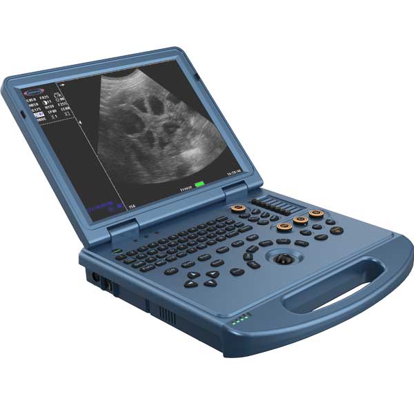
- System: portable veterinary color Doppler ultrasound (e.g., L3-VET-class system)
- Probe: micro-convex (better for penetration and wider view in large fish)
- Note: for smaller sturgeon, switch to a linear probe for better superficial resolution
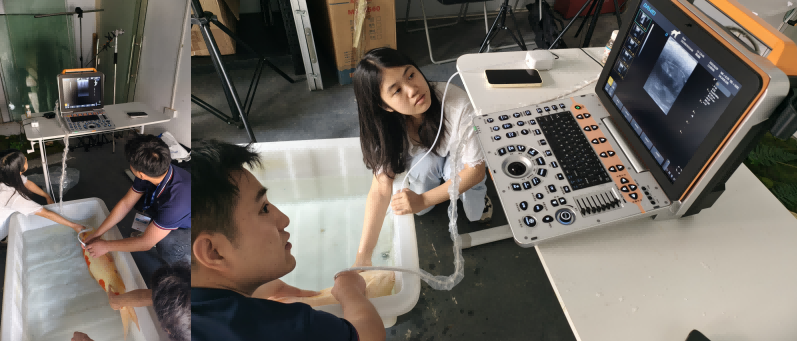
On-site case narrative
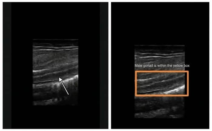
- Fish ultrasound examinations typically include roe assessment, milt assessment, swim bladder checks, and gonad evaluation. In this case, a portable laptop animal ultrasound machine
L3 was used at a sturgeon farm in Hubei to assess gonads and roe. A micro-convex probe was selected for the large sturgeon; for smaller individuals, a linear probe can be used.
Customer benefits
- Higher efficiency in broodstock screening with fewer invasive checks
- Traceable imaging records to support breeding plan management
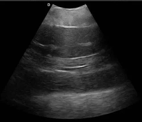
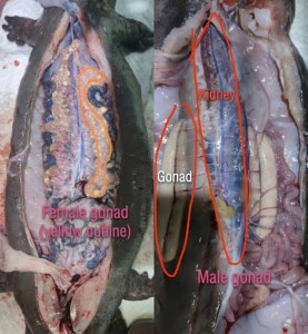
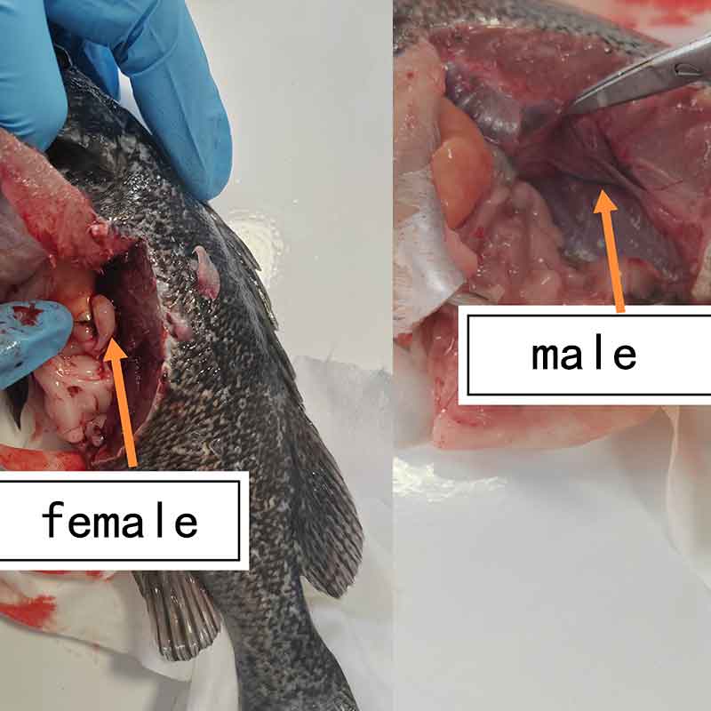
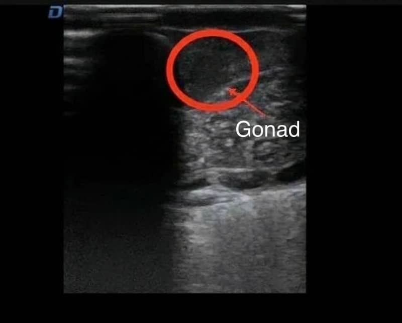
Male gonad ultrasound examination
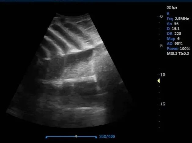
Female gonad examination
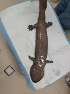
2.Chinese Giant Salamander
A research institute in Yunnan called, expressing their interest in having Dawei Veterinary Medical demonstrate the use of the L3-VET laptop-style veterinary color Doppler ultrasound for examining the gonads of Chinese giant salamanders. Since the animals are relatively small, we will use the linear array probe we provide.
3. Black Grass Carp Ultrasound
Objective
- Gonad examination for breeding management (gonad ultrasound, ultrasound sexing fish)
System & Probe Setup
- Ultrasound system: Dawei P60-VET Portable Veterinary Color Doppler Ultrasound
- Probe: High-frequency linear transducer (recommended for clearer superficial-detail imaging)
Application Note
- For black rockfish, pairing the P60-VET with a high-frequency linear probe enables better visualization of gonadal structures, improving on-site sexing and reproductive assessment efficiency.
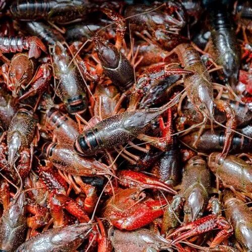
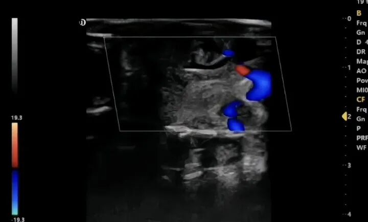
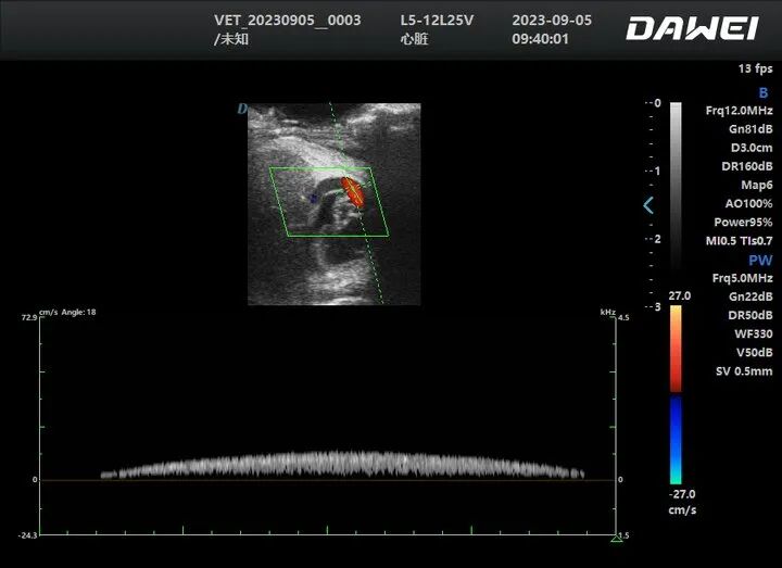
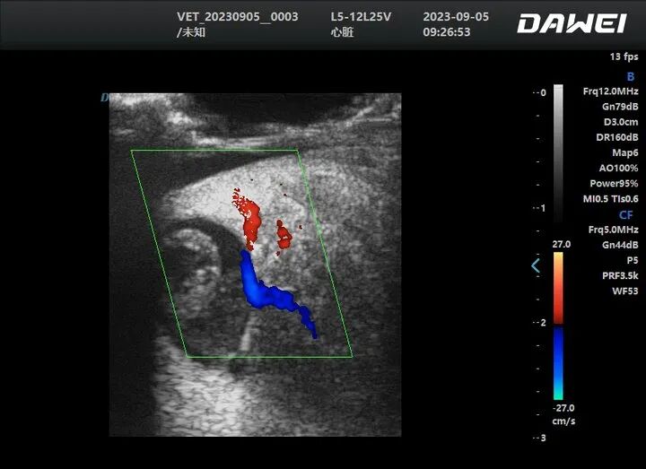
4. Crayfish Ultrasound Case
Objective
- Monitor crayfish cardiac activity and heart rate changes to assess how water temperature, water quality, and other environmental factors affect growth.
System
- Dawei P5-VET Portable Veterinary Color Doppler Ultrasound
How it’s used
- Pond/lab scanning enables real-time heart rate trend recording, then results are compared across different environmental conditions.
Value
- Turns physiological signals into actionable data, helping research teams and farms move from experience-based decisions to data-driven management.
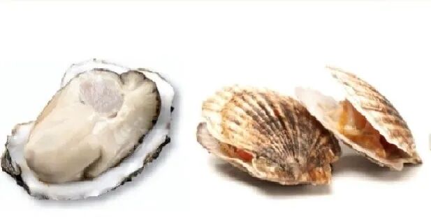
5. Shellfish ultrasound / bivalve ultrasound Case
Objective
- Measure heart rate changes in oysters and scallops under different water temperature conditions, to support aquaculture research and provide practical guidance for farmers.
System
- Dawei L5-VET Laptop Veterinary Color Doppler Ultrasound
- Probe: High-frequency linear transducer (for better superficial-detail imaging)
How it’s used
- Researchers perform ultrasound scanning and record heart rate signals at different temperature settings, then compare trends across groups.
Value
- Converts biological signals into quantifiable data, helping develop science-based temperature management recommendations for shellfish farming.
6. Koi ultrasound Case
Objectives
- Breeding management: gonad development assessment, grouping and pairing support
- Body condition and abdominal structure observation for daily management and show-level care (not a substitute for veterinary diagnosis)
Solution setup (recommended)
- Large koi: micro-convex / convex
- Small–medium koi: linear
- Suggested accessories: protective cover + rapid disinfection workflow for high-frequency use
Customer benefits
- Faster breeding decisions and improved management precision
Fish Solution Overview(System + Probes + Workflow)
Dawei Veterinary Medical provides a wide range of veterinary ultrasound models with a complete selection of probes. In addition to the aquatic species mentioned above, our systems can also be used for ultrasound examination in turbot, salmon, stingrays, trout, and more.
Recommended fish ultrasound system form factor
- Handheld B/W ultrasound、Portable veterinary color Doppler ultrasound for pond-side / hatchery / on-boat use
- Outdoor power options: internal battery, external battery pack, or vehicle power
- Easy cleaning & protection: durable housing and wipe-down disinfection compatibility (as applicable)
Probe selection guideline (by size & anatomy)
General logic below; final selection should match your probe models and frequency ranges.
| Target size / anatomy | Preferred probe | Typical advantage | Notes |
| Large fish (sturgeon, large koi): abdomen & gonads | Micro-convex / convex | Better penetration, wider field of view | Often easier pond-side |
| Small–medium fish: abdomen & gonads | Linear | Higher resolution for superficial structures | Best for smaller bodies |
| Shellfish / superficial structures (some bivalves) | High-frequency linear | Better near-field detail | Usually needs customized coupling & fixation |
Suggested on-site SOP (workflow)
- Capture & short restraint: minimize pressure and handling time
- Surface prep & coupling: manage mucus/water film and choose proper coupling method
- Scan route: ventral/lateral views; record by zones (front–mid–rear)
- Save key images: B-mode + Color (if needed) + measurements (if needed)
- Interpretation & grouping: build a traceable log (image–ID–time–weight–notes)