


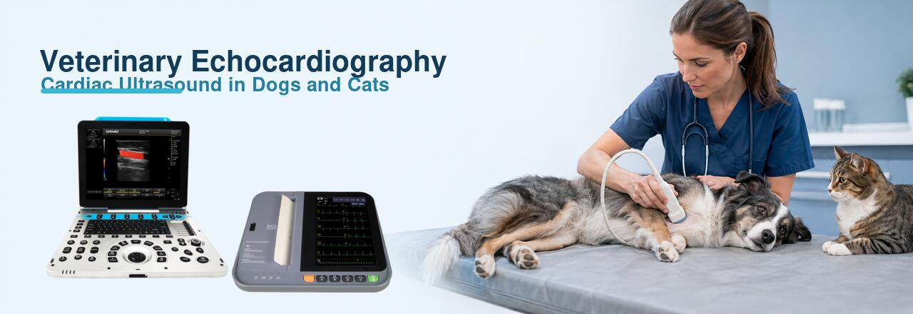
Veterinary Echocardiography: How to Perform Cardiac Ultrasound in Dogs and Cats
Veterinary echocardiography is a non-invasive ultrasound examination used to evaluate the structure, movement and blood flow of the heart. In dogs and cats, it can help veterinarians assess chamber size, myocardial thickness, valve motion, cardiac contractility and abnormal blood flow in real time.
A complete cardiac evaluation often combines echocardiography with an electrocardiogram. Ultrasound shows how the heart is built and how it moves, while ECG records its electrical activity. Together, the two examinations provide a more complete picture of cardiac health than either test alone.
Quick Answer: How Is a Veterinary Echocardiogram Performed?
A veterinary echocardiogram is usually performed with the dog or cat positioned in right and left lateral recumbency. After a small area of hair is clipped, ultrasound gel is applied and a phased-array cardiac probe is placed over the parasternal acoustic windows.
The examination normally includes:
- Right parasternal long-axis views
- Right parasternal short-axis views
- Left apical views
- Left cranial views
- Two-dimensional and M-mode imaging
- Color, pulsed-wave and continuous-wave Doppler
- Selected cardiac measurements and ECG recording
Standardized imaging planes make examinations easier to repeat, compare and report. The recognized transthoracic approach uses right parasternal, left apical and left cranial acoustic windows. This is consistent with established veterinary echocardiography standards for dogs and cats.
When Do Dogs and Cats Need Cardiac Ultrasound?
A veterinarian may recommend echocardiography when a physical examination or another diagnostic test suggests possible heart disease.
Common indications include:
- Heart murmur
- Irregular heart rhythm
- Persistent coughing or breathing difficulty
- Exercise intolerance
- Weakness, collapse or fainting
- Abnormal heart size on radiographs
- Suspected cardiomyopathy
- Suspected congenital heart disease
- Pre-anesthetic cardiac assessment
- Follow-up of previously diagnosed heart disease
In cats, echocardiography is particularly important when hypertrophic cardiomyopathy is suspected. Some affected cats may not have an obvious heart murmur, so imaging is often needed to assess ventricular wall thickness, chamber size and blood flow.
Cardiac ultrasound should be interpreted alongside the animal’s clinical history, physical examination, blood pressure, thoracic radiographs, laboratory results and ECG findings.
Choosing an Ultrasound System for Veterinary Echocardiography
Not every ultrasound machine is equally suited to cardiac examinations. The heart moves rapidly, especially in cats and small dogs. Good temporal resolution, stable Doppler performance and an appropriate cardiac probe are therefore essential.
A suitable veterinary color Doppler ultrasound system should provide:
- High-quality two-dimensional imaging
- M-mode
- Color flow Doppler
- Pulsed-wave Doppler
- Continuous-wave Doppler for high-velocity flow
- Adjustable sweep speed
- Cardiac measurement packages
- Cine-loop storage
- A phased-array cardiac probe
- ECG synchronization where available
A portable system can suit mobile services, general veterinary clinics and examination rooms with limited space. A trolley system is often better suited to animal hospitals that perform frequent or advanced cardiac examinations and require a larger display, multiple probe ports and a more complete workflow.
Preparing the Patient
Most dogs and cats can undergo echocardiography without general anesthesia. Sedation is not routinely required because some sedatives can alter heart rate, contractility and blood pressure. However, the veterinarian may consider mild sedation when stress or movement makes examination impossible.
Cats should be handled gently in a quiet environment with minimal restraint. Many cats can be examined in lateral recumbency, although standing or sternal positioning may be used when respiratory distress prevents conventional positioning.
Before scanning:
- Confirm the animal’s identity, weight and clinical history.
- Record heart rate, respiratory rate and relevant physical findings.
- Clip a small area over the right and left cardiac windows when necessary.
- Apply ultrasound gel to remove air between the probe and skin.
- Attach ECG electrodes when simultaneous rhythm monitoring is required.
- Allow the patient to settle before recording measurements.
Excessive pressure from the probe should be avoided, particularly in cats and patients experiencing respiratory difficulty.
Step 1: Obtain the Right Parasternal Long-Axis Views
Begin with the patient in right lateral recumbency. Place the phased-array probe near the right parasternal cardiac window and adjust its position until the heart is clearly visualized.
Four-Chamber View
The right parasternal four-chamber view displays the:
- Right atrium
- Right ventricle
- Left atrium
- Left ventricle
- Mitral valve
- Tricuspid valve
This view allows the operator to examine chamber proportions, atrioventricular valve motion, ventricular wall movement and pericardial space.
Depth should be reduced enough to fill the screen with the heart while keeping the entire cardiac structure visible. Gain should reveal the endocardial borders without filling the chambers with excessive noise.
Left Ventricular Outflow View
A slight rotation or angulation of the probe brings the left ventricular outflow tract, aortic valve and ascending aorta into view.
This image is useful for evaluating:
- Aortic valve morphology
- Left ventricular outflow obstruction
- Subaortic abnormalities
- Aortic root dimensions
- Color flow through the outflow tract
When color Doppler is activated, keep the color box as small as practical to maintain frame rate.
Step 2: Obtain the Right Parasternal Short-Axis Views
Rotate the probe approximately 90 degrees from the long-axis orientation. By gradually angling the beam from the cardiac apex toward the base, several standard short-axis levels can be obtained.
Important levels include:
- Left ventricular apex
- Papillary muscles
- Chordae tendineae
- Mitral valve
- Aortic valve
- Pulmonary artery
At the papillary muscle level, the left ventricle should appear circular rather than oval. This is commonly used for two-dimensional and M-mode assessment of left ventricular size and motion.
At the heart-base level, the aortic valve is centrally positioned, with the right-sided cardiac structures surrounding it. This view is commonly used to assess the left atrium-to-aorta relationship and the morphology of the right heart.
Step 3: Use M-Mode for Motion and Measurements
M-mode records the movement of cardiac structures along a single ultrasound line over time. It offers high temporal resolution and is helpful when measuring rapidly moving structures.
Common uses include:
- Left ventricular internal dimensions
- Interventricular septal thickness
- Left ventricular free-wall thickness
- Fractional shortening
- Mitral valve motion
- Heart rate and motion timing
The M-mode cursor should be guided by a correctly aligned two-dimensional image. An oblique cursor can overestimate wall thickness or chamber dimensions.
For cats suspected of cardiomyopathy, measurements should not rely on only one M-mode image. Focal hypertrophy may be missed, so wall thickness should also be assessed in multiple two-dimensional views. The ACVIM feline cardiomyopathy guidance recommends evaluating wall thickness across more than one cardiac view and averaging measurements from multiple cardiac cycles.
Step 4: Obtain the Left Apical Views
Move the patient into left lateral recumbency and position the probe over the left caudal or apical cardiac window.
The left apical four-chamber view is especially useful for Doppler assessment because it helps align the ultrasound beam with blood flow through the mitral and tricuspid valves.
From this window, the operator can obtain:
- Left apical four-chamber view
- Left apical two-chamber view
- Left apical five-chamber view
- Mitral inflow Doppler
- Aortic outflow Doppler
- Tissue Doppler measurements, when supported
Good Doppler measurements depend on alignment. The ultrasound beam should be as parallel to blood flow as possible. Even a moderate angle error can underestimate flow velocity.
Step 5: Evaluate Blood Flow with Doppler Ultrasound
Veterinary color Doppler adds information that cannot be obtained from a grayscale image alone.
Color Doppler
Color Doppler shows the direction and distribution of blood flow. It can help identify:
- Mitral or tricuspid regurgitation
- Aortic or pulmonary insufficiency
- Abnormal intracardiac communications
- Turbulent flow
- Outflow tract obstruction
Color alone does not determine the exact severity of disease. Findings should be confirmed with spectral Doppler and interpreted with the rest of the echocardiographic examination.
Pulsed-Wave Doppler
Pulsed-wave Doppler measures blood-flow velocity at a selected sample location. It is useful for assessing lower-velocity flow through valves and vessels and for evaluating filling patterns.
Continuous-Wave Doppler
Continuous-wave Doppler records high velocities along the entire ultrasound beam. It is particularly useful when evaluating high-speed jets caused by stenosis, obstruction or significant regurgitation.
If you are comparing the roles of grayscale and flow imaging, see B-Mode vs Doppler Ultrasound for Animal Diagnosis.
Step 6: Record a Veterinary ECG
Echocardiography and ECG answer different clinical questions.
An echocardiogram evaluates:
- Cardiac anatomy
- Chamber dimensions
- Wall thickness
- Valve motion
- Contractility
- Blood-flow direction and velocity
An electrocardiogram evaluates:
- Heart rhythm
- Heart rate
- Electrical conduction
- Arrhythmias
- Timing of cardiac electrical events
When an irregular rhythm is observed during ultrasound, a diagnostic ECG can help determine whether it represents atrial fibrillation, premature complexes, conduction abnormalities or another rhythm disturbance.
The Dawei veterinary ECG product range includes systems for different clinical requirements. The DE12A-VET 12-channel veterinary ECG machine features a 10.2-inch color touchscreen, synchronized ECG acquisition, multiple recording and display modes, automatic measurement and analysis, waveform freezing and storage of up to 1,000 records.
Veterinarians can view the detailed configuration on the DE12A-VET 12-channel veterinary ECG machine page.
An automatic ECG interpretation should be treated as a clinical reference rather than a replacement for professional evaluation.
Dog and Cat Echocardiography: Important Differences
The basic scanning windows are similar, but the examination must be adapted to the patient.
Dogs vary greatly in body size and chest shape. Probe frequency, depth and sector width may need substantial adjustment between a small breed and a large dog.
Cats are smaller and normally have faster heart rates. Higher-frequency phased-array probes, shallow imaging depth, narrow sector width and high frame rates are especially valuable. Gentle handling is also critical because stress can rapidly increase feline heart rate and interfere with Doppler measurements.
Reference values must be selected according to species, body size and measurement method. Canine measurements often require body-weight normalization, while feline wall thickness and atrial dimensions must be interpreted in the context of the complete examination.
Common Echocardiography Errors
Using the Wrong Probe
A large convex abdominal probe may not fit between the ribs or provide adequate cardiac resolution. A phased-array probe with a small footprint is generally preferred.
Measuring an Oblique Image
An incorrectly aligned view can make chambers or myocardial walls appear larger than they are. Measurements should be taken only after a standard imaging plane has been obtained.
Using Too Much Imaging Depth
Excessive depth reduces the size of the heart on the screen and may lower frame rate. Set the depth just beyond the cardiac structure of interest.
Making the Color Box Too Large
A large color Doppler box can reduce temporal resolution. Limit the box to the valve or vessel being examined.
Relying on One Cardiac Cycle
Respiration, movement and rhythm variation can change measurements. Record representative cine loops and average multiple cycles when appropriate.
Treating Focused Ultrasound as a Complete Echocardiogram
Focused cardiac ultrasound can support rapid triage, but it does not replace a complete standardized echocardiographic examination when detailed diagnosis is required.
Choosing Between Portable and Trolley Veterinary Ultrasound
The right system depends on examination volume and clinical workflow.
The P3-VET portable veterinary color Doppler ultrasound is designed for applications that include small-animal cardiac function and blood-flow assessment. It supports two-dimensional imaging, M-mode, color Doppler, PW, CW and tissue Doppler functions. Its portable format is suitable for clinics that need flexibility between examination rooms or mobile services.
For animal hospitals performing frequent ultrasound examinations, the T8-VET trolley veterinary ultrasound provides a 21.5-inch medical display, adjustable touchscreen, multiple probe ports and optional phased-array cardiac probe support. Its trolley configuration is suitable for a fixed imaging room and a higher-volume clinical workflow.
Veterinary Echocardiography Reporting Checklist
A structured report should include:
- Patient identification and body weight
- Clinical indication
- Heart rhythm and heart rate
- Image quality
- Chamber dimensions
- Myocardial thickness
- Valve morphology and motion
- Systolic function
- Color and spectral Doppler findings
- Pericardial or pleural abnormalities
- Main interpretation
- Recommended follow-up or referral
Images and cine loops should be stored with clear labels so that future examinations can be compared consistently.
Frequently Asked Questions
Can a regular veterinary ultrasound machine perform echocardiography?
Only if it has suitable cardiac imaging functions and an appropriate probe. A system intended mainly for abdominal or pregnancy examinations may lack the phased-array probe, high frame rate, CW Doppler or cardiac measurement package needed for detailed echocardiography.
Which probe is best for dog and cat heart ultrasound?
A phased-array cardiac probe is generally preferred because its small footprint fits between the ribs while producing a sector-shaped image of the heart. Probe frequency should be selected according to the animal’s size and imaging depth.
Is sedation necessary for a veterinary echocardiogram?
Most dogs and cats can be examined without sedation. When sedation is necessary, the drug and dose should be selected by the veterinarian because medication may affect cardiovascular measurements.
What is the difference between an ECG and an echocardiogram?
An ECG records the electrical activity and rhythm of the heart. An echocardiogram uses ultrasound to examine cardiac structure, motion and blood flow. The two examinations complement rather than replace each other.
How long does a dog or cat echocardiogram take?
The duration depends on patient cooperation, disease complexity and whether a focused or complete examination is required. A complete study generally takes longer than a rapid point-of-care cardiac scan.
Can echocardiography diagnose every heart condition?
No. Echocardiography is highly valuable but operator-dependent. Some conditions require ECG, radiographs, blood pressure measurement, cardiac biomarkers or other diagnostic tests. Difficult or complex cases may require assessment by a veterinary cardiologist.
Conclusion
Veterinary echocardiography provides real-time information about cardiac anatomy, movement and blood flow in dogs and cats. A reliable examination begins with appropriate patient preparation, a phased-array probe, standardized imaging windows and correct use of two-dimensional, M-mode and Doppler imaging.
Combining cardiac ultrasound with a veterinary ECG helps connect structural findings with electrical activity. With suitable equipment, standardized technique and professional interpretation, veterinary clinics can build a more complete and repeatable cardiac diagnostic workflow.
Post time: Aug-13-2026