


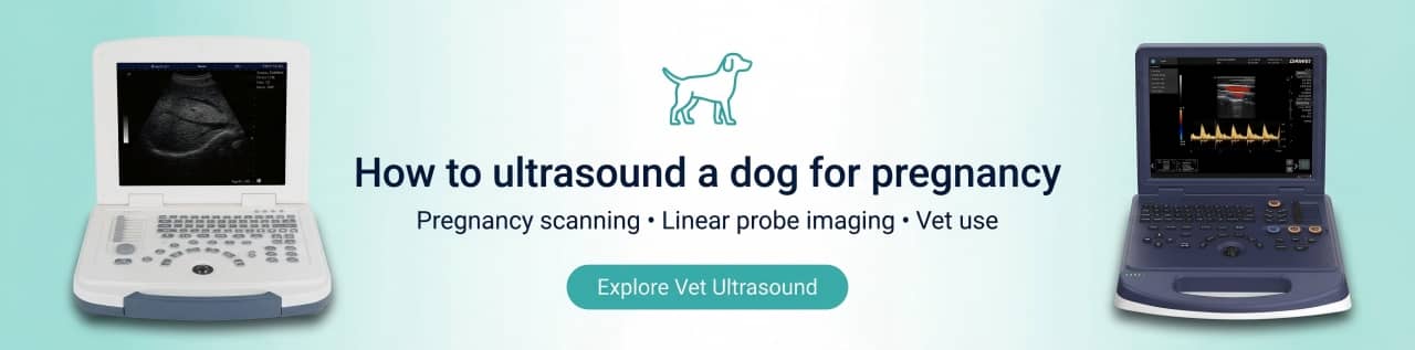
Ultrasound scanning to diagnose early pregnancy in dogs can detect the embryonic sac as early as 20d after mating, the accuracy of early pregnancy diagnosis in dogs: dogs in the 21-25d after mating can reach 86%. Using vet ultrasound systems to detect dog pregnancy method, guide animal reproduction, the method is safe, reliable and convenient.
Why ultrasound is used for canine pregnancy
Ultrasound is widely used in veterinary reproduction because it is non-invasive, does not use ionizing radiation, and can help a veterinarian:
- Confirm pregnancy earlier than X-ray
- Assess fetal viability (e.g., fetal heart activity)
- Estimate gestational age using measurements (when performed by trained professionals)
- Differentiate pregnancy from conditions such as pyometra, hydrometra, or uterine enlargement
Evidence-based timing notes :
- Merck Veterinary Manual notes ultrasonography is best performed at 25–35 days gestation and false-negative results may occur before 21 days.
- A Theriogenology review (Root Kustritz, 2005) reports amniotic vesicles may be visible as early as day 19–20, but most authors recommend scanning no earlier than day 25 to reduce missed diagnoses.
So how to do ultrasound examination for pregnant dogs?
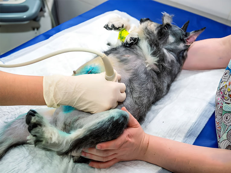
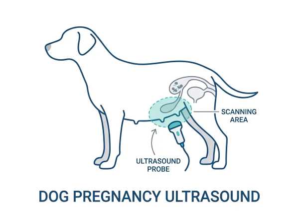
1. Place the bitch on a comfortable examination table or in a quiet room in the veterinarian's office. The probing area is at the rear helpers, the edge of the udder. If the dog's hair is long, it still needs to be clipped, if not, just separate the hair.
2. The veterinarian will apply some coupling agent to the skin of the dog's abdomen to help the ultrasound probe slide better and transmit sound waves.
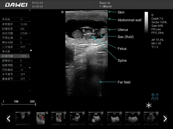
3. To perform ultrasound on a dog, a 6.5MHz microconvex probe may be used to start at the pubic symphysis, detect the bladder and then probe forward to the uterine region on both sides, moving the probe as it goes in order to obtain images at different angles.
4. The veterinarian will observe the images on the screen to determine if a fetus is present, as well as the number, size and location of the fetuses.
5. The veterinarian will use the ultrasound images to confirm the health status of the puppy. If needed, the veterinarian can also measure the heart rate of the fetuses to make sure they are healthy.
Dog ultrasound scanning is a very safe and painless method of providing veterinarians and dog owners with critical information about the health of the bitch and fetus. dog pregnancy ultrasound week by week is very important to maintain your dog's pregnancy health. so Canine ultrasound scans used to determine pregnancy are an important tool to ensure the health of pregnant dogs and their future puppies.To learn more about canine veterinary ultrasound systems, come and ask us.
When to ultrasound a dog for pregnancy (timing guide)
Because dogs have a relatively short gestation (often ~63 days on average), timing matters. Here’s a practical vet-style timing framework breeders commonly use.
Day-by-day highlights
- Day 19–22: On high-quality equipment, the earliest fluid-filled gestational structures may sometimes be seen, but intestinal gas and small size can cause false negatives.
- Day 25–35: Commonly recommended window to confirm pregnancy and evaluate fetal viability.
- After ~day 42–45: X-ray becomes more useful for counting puppies (fetal skeleton mineralization becomes visible).
Pregnancy ultrasound week by week (what is realistic)
| Week (approx.) | Days after breeding | What ultrasound can help with | Notes |
| Week 3 | 19–21 | Possible early pregnancy signs | Higher miss rate; avoid over-promising results |
| Week 4 | 22–28 | More reliable confirmation; early heart activity may be detectable | Best handled by experienced operators |
| Week 5 | 29–35 | Strong confirmation; viability checks; early estimates | Counting is still unreliable |
| Week 6–7 | 36–49 | Monitoring growth/viability; spotting problems | Counting remains difficult |
| Week 8–9 | 50–63 | Late checks; consider X-ray for litter size | Plan whelping support |
What equipment is used for dog pregnancy ultrasound?
Veterinarians typically use B‑mode (real-time) ultrasound for canine pregnancy diagnosis.For dog pregnancy ultrasound, clinics often match the system to their workflow—some prefer a basic laptop veterinary ultrasound
(e.g., MU10), others need a portable veterinary ultrasound scanner for on-the-go use (e.g., Slite), and some choose an entry-level color Doppler pet ultrasound system (e.g., L30i) for broader imaging needs.
Probe selection (typical ranges)
- Many dogs: ~5.0 MHz transducer can work well (general reference in veterinary literature).
- Toy breeds / very small dogs: higher frequency (e.g., ~7.5 MHz) may improve resolution.
- Microconvex probes are popular for small-animal abdominal scanning because they are easier to position.
Imaging features that help
If you’re comparing systems for clinics or breeders, common feature keywords that matter for dog pregnancy scanning include:
- Portable veterinary ultrasound machine (field use)
- Microconvex probe / abdominal probe
- B‑mode, M‑mode for heart activity checks
- Image optimization: gain, depth, TGC, focus
- Freeze/zoom and measurement packages (GSD/CRL/BPD)
Preparation checklist (before the scan)
A clean, calm setup improves image quality and reduces stress:
- Quiet room + non-slip exam table
- Clip or part long hair on the lower abdomen
- Use adequate coupling gel
- Gentle restraint (avoid compressing the abdomen too firmly)
- If the dog is anxious, discuss calm-handling strategies with a veterinarian
Some veterinary guidance suggests keeping the bladder as a landmark; follow your clinic’s protocols.
How veterinarians scan: positioning & technique (expanded)
Below is a more detailed (but still practical) technique overview that matches common clinical workflows.
Common positions
- Dorsal recumbency (on the back) is common for abdominal reproductive scans.
- Lateral recumbency (on the side) may be used if the dog is uncomfortable on her back.
A simple scanning route (conceptual)
- Identify the bladder as a caudal landmark.
- Sweep cranially and laterally to evaluate both uterine horns.
- Use multiple angles (fan/rock/slide) to avoid mistaking bowel gas for pregnancy structures.
- If viability is being checked, confirm fetal heart activity using the machine’s appropriate mode/settings.
Interpreting results: what ultrasound can and can’t tell you
What it can tell you (best use)
- Pregnancy confirmation in the recommended window
- Viability indicators (e.g., fetal heart activity)
- General uterine assessment when pregnancy is uncertain
- Accurate puppy count: even good scans can miss fetuses, especially with large litters or late gestation. If an exact count is needed, veterinarians often recommend radiography in late pregnancy.
What it cannot reliably do
- Accurate puppy count: even good scans can miss fetuses, especially with large litters or late gestation. If an exact count is needed, veterinarians often recommend radiography in late pregnancy.
Common Problems in Pet Ultrasound (FAQ)
How soon can a dog pregnancy be detected by ultrasound?
Sometimes as early as ~day 20, but many veterinary sources recommend ~day 25+ for a more reliable scan.
Is dog ultrasound safe during pregnancy?
Diagnostic veterinary ultrasound is generally considered safe and non-invasive when performed correctly.
Can I do a pregnancy ultrasound at home?
Home scanning without training can cause misinterpretation and unnecessary stress. For pregnancy confirmation and health decisions, consult a veterinary professional.
What’s the best probe for dog pregnancy ultrasound?
Many clinics use a microconvex abdominal probe in the ~5–8 MHz range (choice varies by dog size and machine).
With DAWEI Veterinary Medical’s basic laptop veterinary ultrasound MU10 paired with a 5.0 MHz micro-convex probe, you can achieve fast, accurate, real-time ultrasound scanning for dogs.
This MU10 + 5.0 MHz micro-convex setup is also a great option for abdominal ultrasound examinations in sheep, cats, and rabbits.
Post time: Sep-21-2023