


Black & White Veterinary Ultrasound Diagnostic System Pseudo Color Processing is a color mapping process whose main purpose is to enhance the veterinarian’s ability to detect information in veterinary ultrasound images.Black & white veterinary ultrasound Pseudo Color Technology is a visualization method commonly used in animal diagnostic medical ultrasound imaging that uses pseudo-color coding to enhance the contrast of the image, making it easier to distinguish between different tissues and lesions. This technique is very useful in animal medical diagnostics because it helps veterinarians more accurately identify and analyze structures and problems within the animal’s body.
How black & white veterinary ultrasound diagnostic system pseudo-color technology works:
Data Acquisition: When performing animal ultrasound imaging , an ultrasound probe emits high-frequency sound waves into the animal’s body and then receives the returned echo signals. These signals are processed and converted into an image, resulting in a black and white ultrasound image.
Gray Scale Mapping: Conventional black and white ultrasound images represent the intensity and reflectivity of different tissues as gray scale values. The reflectivity of sound waves from different tissues results in variations in brightness in the image. However, it is not always intuitive and easy for physicians to differentiate between different tissues and lesions simply by using grayscale images.
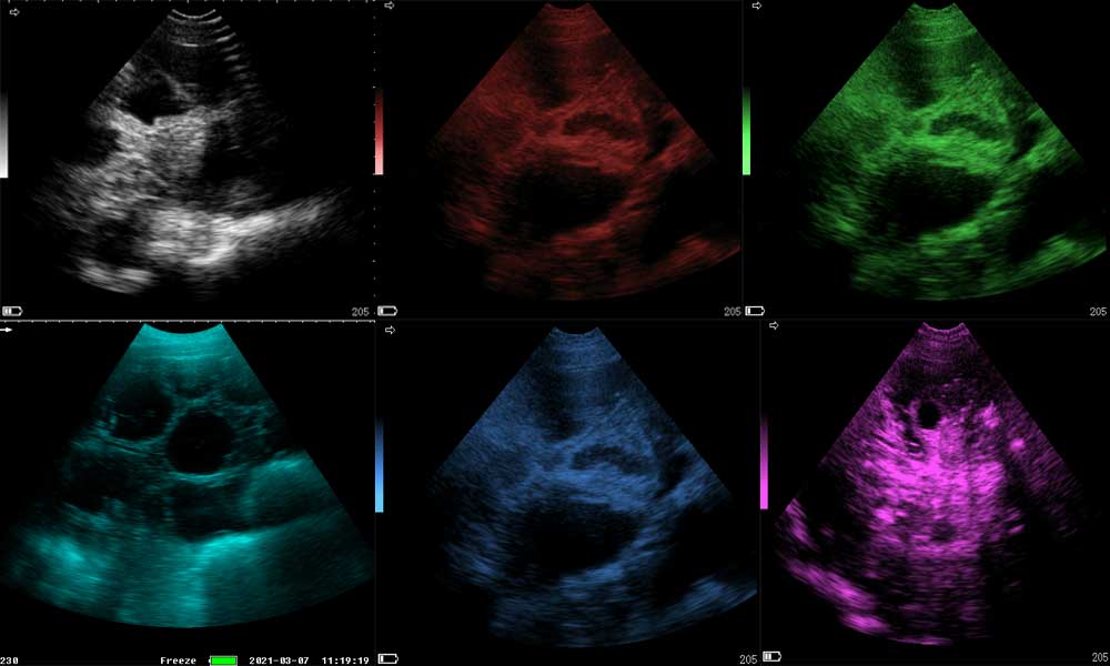
Pseudo-color coding: In order to enhance image visualization, pseudo-color coding techniques are applied to black-and-white ultrasound images. This coding makes it easier for physicians to distinguish between different tissue types or lesions by mapping different grayscale values to different colors. Common pseudo-color coding schemes include mapping low gray values to dark blue or black, middle gray values to green, and high gray values to red.
Veterinary ultrasound Image Display: Once pseudo-color coding has been applied, the image will appear in color on the monitor. Different tissues or lesions will be displayed in different colors, making it easier for the doctor to identify and understand the information in the image.
Dawei Veterinary B/W Ultra Diagnostic System Pseudo-color Processing in Gray Scale – Color Transformation Method is a representative method in Veterinary B/W Ultra Pseudo-color Processing, which can change the black and white grayscale image into a continuous color image with multiple color gradients. It can change the black and white grayscale image into a continuous color image with multiple color gradients. The color it maps, as far as the current level is concerned, generally up to 512 kinds of more than pseudo-color veterinary black and white ultra-transformation of the image visual effect is good, in the veterinary clinical diagnosis of more than a number of applications.
In short, in the black & white vet ultrasound diagnostic System instrument color process, combined with clinical needs, flexible and appropriate use of the above technology, can improve the veterinary ultrasound image can be identified, thereby improving the diagnosis rate.
Post time: Aug-14-2023