


An ultrasound machine can have excellent imaging hardware and still be the wrong choice for a veterinary practice if it cannot support the probes that practice actually needs.
That point is easy to miss. Buyers often compare screens, Doppler modes, battery life and software first. Yet the transducer is the part that physically meets the animal. Its footprint determines where you can scan. Its array geometry changes the field of view. Its frequency range influences the trade-off between penetration and detail. And in cattle, horses or production animals, the probe design can even determine the route and purpose of the examination.
So, what are the main veterinary ultrasound probe types, and when should each one be used?
Quick answer: which veterinary ultrasound probe is used for what?
For most veterinary buyers, the fastest way to understand the options is to start with the examination rather than the probe name:
- Convex probe: broad abdominal and reproductive surveys where useful depth and a wide field are priorities.
- Microconvex probe: small-animal abdominal and thoracic work where a compact footprint must still provide a widening field of view.
- Linear probe: superficial structures such as tendons, muscles, vessels and small parts where near-field detail matters.
- Phased array probe: echocardiography and other examinations that need a small intercostal footprint and a sector-shaped image.
- Rectal linear or rectal convex probe: transrectal reproductive examinations in cattle and horses.
- Backfat probe: specialized body-composition measurements, especially backfat and loin/eye-muscle assessment in swine production workflows.
There is overlap. That is normal. A microconvex and a convex probe can both image an abdomen; a linear and microconvex probe may both be useful in thoracic point-of-care ultrasound. The better question is not “Which probe is best?” It is “Which probe gives me the right access, depth and image geometry for this patient and this exam?”
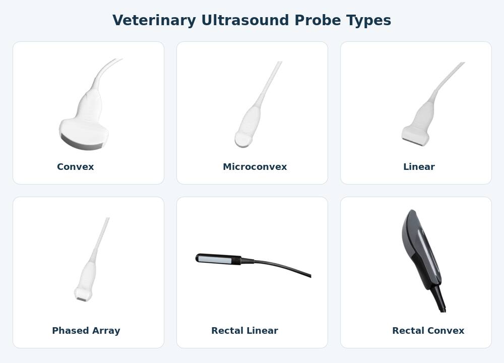
Figure 1. Four common general diagnostic veterinary probe types using Dawei’s own probe assets.
Veterinary ultrasound probe types at a glance
| Probe type | Typical image / footprint | Common veterinary applications | Main advantage | Key limitation |
|---|---|---|---|---|
| Convex | Curved, relatively broad footprint; widening field | Abdomen, reproduction, deeper targets in medium and large animals | Depth plus broad survey view | Footprint can be large for tiny patients or narrow rib spaces |
| Microconvex | Small curved footprint; widening field | Cats, small dogs, small-animal abdomen, tight acoustic windows | Compact contact area with useful depth | Less superficial detail than a high-frequency linear probe in many exams |
| Linear | Flat footprint; rectangular field | Tendons, ligaments, vessels, superficial masses, guided procedures | Excellent near-field detail | Higher-frequency settings sacrifice penetration |
| Phased array | Very small footprint; sector field | Echocardiography, intercostal cardiac windows | Small contact area with a field that opens at depth | Less suitable for broad superficial imaging |
| Rectal linear / convex | Elongated transrectal design; geometry varies by array | Cattle and equine reproductive examinations | Purpose-built access to uterus and ovaries | Specialized route; not a routine external abdominal probe |
| Backfat | Specialized measurement/contact assembly | Swine backfat and loin/eye-muscle measurement | Repeatable production-animal measurement workflow | Purpose-specific rather than general diagnostic scanning |
That table captures the broad differences. The real decision becomes clearer when we look at the probes one by one.
1. Convex probe: the abdominal workhorse
A convex—or curvilinear—probe has a curved acoustic footprint. Its image widens with depth, which is useful when you need to survey a relatively large region of the abdomen without moving the probe constantly.
This geometry makes convex probes a practical choice for routine abdominal and reproductive scanning in many species. They are especially useful when the target lies deeper and penetration matters more than extremely fine superficial detail.
Think about a medium-to-large dog, a sheep, a pig or a bovine abdominal examination. The operator often needs context: liver in relation to surrounding structures, the urinary bladder within the pelvis, or deeper reproductive anatomy. A convex field helps provide that context.
Frequency still matters. Dawei’s livestock ultrasound configurations, for example, include convex options with selectable frequencies rather than one fixed value. That is important because “convex” describes the probe geometry; it does not mean every convex transducer operates at the same frequency.
For large-animal configurations and available transducers, see Dawei’s livestock ultrasound solutions.
2. Microconvex probe: smaller footprint, broad usefulness
A microconvex probe follows the same general idea as a convex transducer, but the contact surface is smaller.
That seemingly modest change can make a large practical difference.
Cats, toy-breed dogs and young animals simply do not offer the same scanning space as a large dog or cow. A compact probe can sit under the costal arch or between ribs with less awkward positioning. Because its field widens beneath the surface, the operator can still see a useful amount of anatomy through that smaller acoustic window.
This is why microconvex probes are popular for small-animal abdominal work. They also appear in thoracic point-of-care ultrasound. A prospective study involving 200 dogs and cats with dyspnea compared microconvex, linear and phased-array transducers for lung ultrasound and found the highest agreement between microconvex and linear probes. The study is a good reminder that more than one transducer may answer the same clinical question, even though their handling and image presentation differ.
For a clinic focused on companion animals, probe compatibility should be considered alongside the console itself. Dawei’s small-animal ultrasound systems are a more relevant starting point than choosing purely by machine size.
3. Linear probe: detail close to the surface
Linear probes use a flat array and produce a rectangular image. They are strongly associated with superficial imaging because many linear transducers operate at relatively high frequencies and give excellent near-field resolution.
In veterinary medicine, this makes them useful for tendons, ligaments, muscle injuries, superficial masses, vascular structures, small parts and ultrasound-guided procedures. Equine tendon scanning is a classic example: the structure of interest is superficial, and fine detail is more valuable than the ability to penetrate deeply into the abdomen.
The trade-off is physics, not marketing. Higher frequency generally improves resolution but attenuates more rapidly as sound travels through tissue. A beautifully detailed image is not helpful if the beam cannot reach the target.
4. Phased array probe: built for cardiac windows
Phased array probes have a very small footprint and create a sector-shaped field that opens with depth. That combination is especially valuable when the operator must work between ribs.
In other words: the physical contact area can be small, yet the heart can still occupy a useful field of view deeper in the thorax.
That is why phased array transducers are closely associated with veterinary echocardiography. Cardiac imaging involves rapidly moving structures, restricted intercostal windows and, often, Doppler functions designed around blood flow and cardiac timing. A phased-array probe is not simply a smaller abdominal probe; its geometry solves a different access problem.
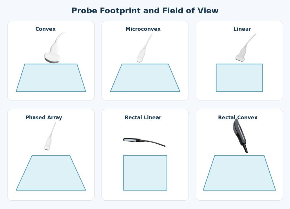
Figure 2. Simplified conceptual comparison of the four general probe geometries. The drawing is illustrative rather than to scale.
5. Rectal probes: essential for many cattle and equine reproductive exams
Now we move beyond the four general external transducers.
For cattle and horses, the examination route can change the probe design entirely. A rectal ultrasound probe is built for transrectal access, allowing the operator to image reproductive structures from a close acoustic window.
Dawei’s probe range includes both a rectal linear design and a separate rectal convex design. They should be treated as two distinct products rather than visually grouped under a generic “rectal probe” label.
Rectal linear probe
The rectal linear probe uses an elongated housing and a linear array. In large-animal reproduction it can be used to evaluate structures such as the uterus and ovaries and to support pregnancy-related examinations, depending on the animal, system and operator technique.
Its form factor matters just as much as its imaging specifications. A standard external linear probe and a rectal linear probe may both use linear-array principles, but they are not interchangeable in handling or intended examination route.
Rectal convex probe
The rectal convex probe is also designed for transrectal use, but its array geometry produces a widening view. This can provide a different field presentation from a rectal linear transducer.
Again, the important point is not that one is universally better. It is compatibility and workflow. A bovine reproduction service should ask which rectal probe the ultrasound platform supports, which frequency options are available and whether that probe matches the examinations routinely performed.
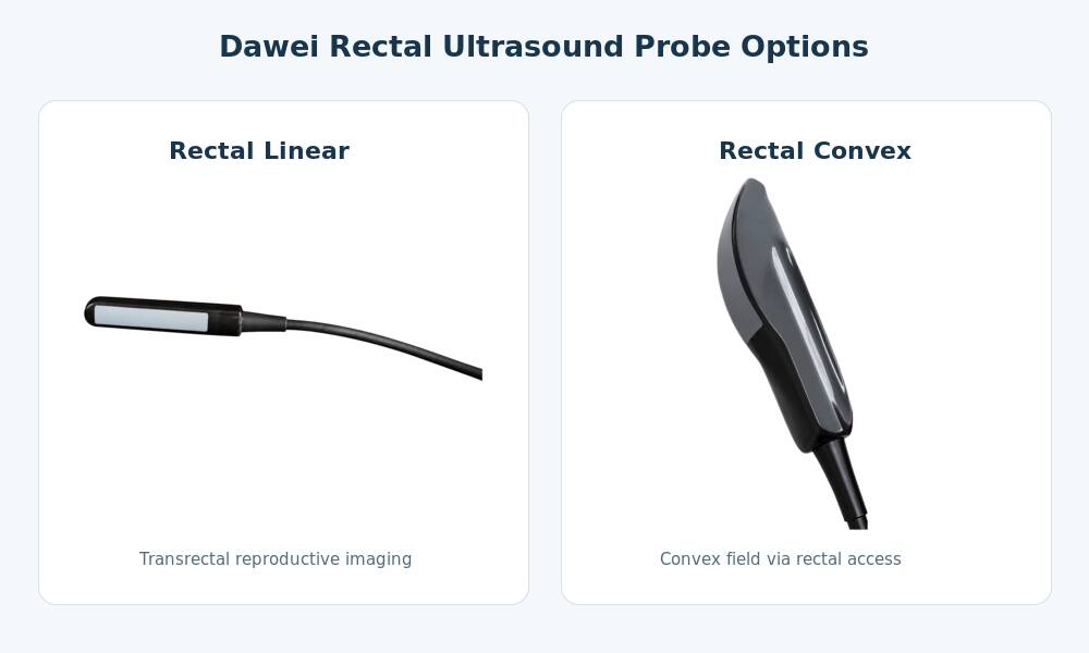
Figure 3. ,shown as separate probe types using the confirmed Dawei product assets.
For additional cattle workflow context, see how veterinarians set ultrasound probe frequency for cattle and using an ultrasound probe for cattle pregnancy examinations.
6. Backfat probe: a specialized tool for production animals
A backfat ultrasound probe belongs in a veterinary probe guide because production-animal ultrasound is not limited to pregnancy diagnosis or disease assessment.
In swine production, ultrasound can also support body-composition measurements. A specialized backfat probe and measurement setup may be used to evaluate subcutaneous fat thickness and, where the system and workflow support it, loin or eye-muscle dimensions.
That is a very different job from general abdominal scanning.
A convex probe is intended to create a diagnostic field through the abdomen. A rectal probe is designed around transrectal reproductive access. A backfat probe is selected because the operator needs a repeatable production measurement. Lumping all three together as “livestock probes” hides the reason they exist.
How probe footprint and frequency work together
Probe selection has two layers.
First, choose a geometry that can physically access the target. Then choose a frequency that can reach it with adequate detail.
In broad terms:
- Higher frequency: better resolution for superficial structures, but less penetration.
- Lower frequency: better penetration for deeper structures, but less fine detail.
Modern broadband probes usually offer selectable operating frequencies, so fixed statements such as “linear equals 7.5 MHz” or “convex equals 3.5 MHz” should be treated cautiously. Those may be useful product examples, not universal definitions.
A more reliable working rule is simple: use the geometry that gives you the correct acoustic window, then use the highest frequency that still reaches the anatomy you need to evaluate. After that, optimize depth, focus and gain.
Which probe should you choose by animal and examination?
Dogs and cats
For routine abdominal work, a microconvex probe is often a practical first choice because its footprint fits small patients and tight acoustic windows. Larger dogs may also be well suited to a standard convex probe. Add a linear probe for superficial structures and a phased array probe if echocardiography is part of the clinical service.
Horses
Probe needs vary dramatically with the examination. Linear probes are important for superficial musculoskeletal work such as tendons. Rectal probes are relevant to reproductive examinations. Convex or other lower-frequency configurations may be considered when deeper imaging is required.
Cattle
For reproductive work, rectal linear or rectal convex probes can become the core transducer rather than an accessory. General external abdominal applications may call for a different probe and frequency range. This is why the phrase “ultrasound for cattle” is not specific enough when choosing equipment: the intended examination must come first.
Sheep and goats
Convex and microconvex probes can both be practical depending on patient size, target depth and scanning technique. The best configuration should be chosen around the actual reproductive or abdominal workflow rather than species name alone.
Pigs
General reproductive or abdominal scanning and backfat measurement are separate workflows. A farm interested in pregnancy detection may need a conventional diagnostic probe configuration, while a breeding or production program interested in body-composition metrics may require a specialized backfat probe.
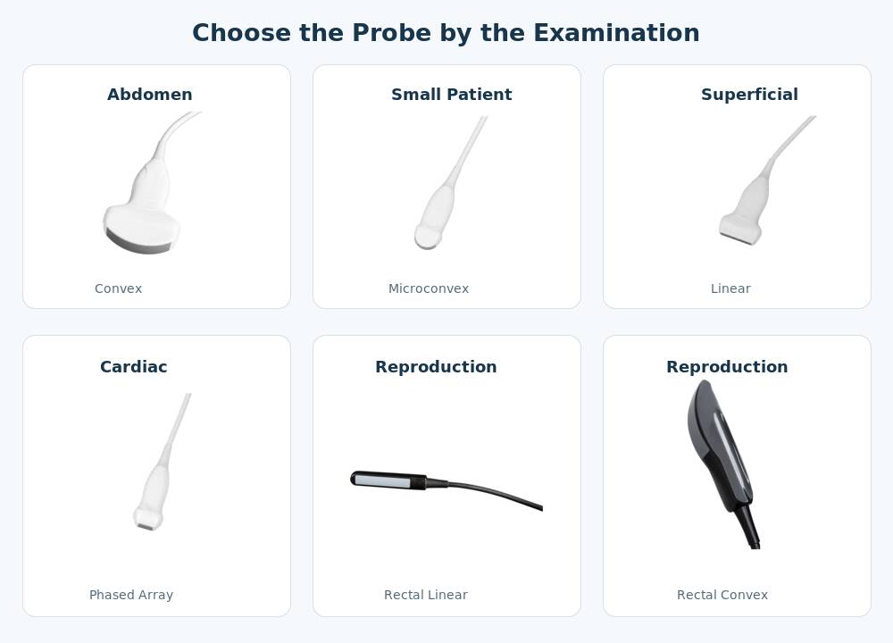
Figure 4. Practical probe-selection starting points. Specialized rectal and backfat workflows should be specified separately when configuring a system.
Microconvex vs convex: how do you decide?
Both produce a widening field. The practical difference begins at the skin.
A standard convex probe gives the operator a larger contact surface and is comfortable for broad abdominal surveys. A microconvex probe reduces that footprint, which can make positioning easier on cats, small dogs or narrow intercostal windows.
If a buyer asks which is “better,” ask about patient size first. The answer may change immediately.
Linear vs phased array: small does not mean similar
These probes can both appear in thoracic workflows, but they prioritize different things.
Linear favors superficial detail and a rectangular image. Phased array favors access through a small intercostal window while opening into a sector at depth.
For a superficial vessel or tendon, linear makes sense. For echocardiography through the rib cage, phased array is usually the more natural choice.
A six-question buying checklist
Before buying a veterinary ultrasound machine—or adding another transducer—answer these questions:
- Which animals will be scanned most often? A feline clinic, equine hospital and pig farm should not start with the same probe bundle.
- What examination is performed most often? Abdomen, heart, tendon, reproductive tract and backfat measurement are different jobs.
- How deep is the target? Depth changes the useful frequency range.
- How large is the acoustic window? Patient size and rib spacing can make footprint geometry decisive.
- Is the examination external or transrectal? Large-animal reproduction may require a purpose-built rectal transducer.
- Which probes are supported by the machine? Never assume that similar-looking probes or connectors are interchangeable across platforms.
This sequence is often more useful than choosing a console first. Define the clinical tasks. Define the required probes. Then shortlist ultrasound systems that support them.
Dawei provides multiple veterinary ultrasound systems covering companion-animal, livestock, equine and reproductive applications. Exact probe availability varies by model and configuration, so the required transducers should be confirmed when requesting a quotation.
Probe care is part of image quality
The acoustic lens, cable, strain relief and connector all experience repeated handling. Drops, tight cable bends, unsuitable disinfectants or poor storage can shorten probe life and introduce artifacts.
Final recommendation
The most useful way to compare veterinary ultrasound probe types is not by memorizing shapes. Match each transducer to the constraint it solves.
Convex gives broad abdominal coverage and depth. Microconvex fits smaller patients and tighter windows. Linear resolves superficial structures. Phased array works through narrow cardiac windows. Rectal probes solve the access problem in cattle and equine reproduction. Backfat probes serve a specialized production-animal measurement workflow.
Once those roles are clear, system selection becomes easier. Instead of asking, “Which veterinary ultrasound machine has the most probes?” ask, “Which machine supports the probes my team will actually use every week?”
That question is far more likely to lead to the right configuration.
FAQ: veterinary ultrasound probe types
What are the main types of veterinary ultrasound probes?
Four common general diagnostic types are convex, microconvex, linear and phased array. Veterinary applications also use specialized rectal linear, rectal convex and backfat probes, particularly in large-animal reproduction and livestock production.
What is the best ultrasound probe for dogs and cats?
There is no single best probe. Microconvex is highly versatile for small-animal abdominal work, linear is useful for superficial structures, and phased array is appropriate for many cardiac examinations. Larger dogs may also be well suited to a standard convex probe.
Which ultrasound probe is used for cattle pregnancy?
Rectal linear and rectal convex probes are purpose-built options for transrectal reproductive examinations in cattle. The correct choice depends on scanning technique and compatibility with the ultrasound system.
Which probe is commonly used for equine tendons?
A linear probe is commonly selected because tendons are superficial structures and benefit from strong near-field detail. Probe frequency should still be adjusted to the required depth and the specific transducer.
Why is phased array used for veterinary echocardiography?
Its small footprint fits between ribs while the sector-shaped image expands at depth, making it well suited to cardiac acoustic windows.
What is a veterinary backfat probe used for?
A backfat probe is used for specialized production-animal measurements such as subcutaneous backfat thickness and, in compatible systems and workflows, loin or eye-muscle dimensions. It is particularly relevant in swine production.
Is a rectal linear probe the same as a standard linear probe?
No. Both may use linear-array principles, but a rectal linear probe has a housing and handling design intended for transrectal examination. A standard external linear probe should not be assumed to serve the same role.
Does a higher-frequency probe always produce a better veterinary ultrasound image?
No. Higher frequency generally improves resolution but reduces penetration. The practical setting is the highest frequency that still reaches the target with adequate image quality.
Can every veterinary ultrasound machine use every probe type?
No. Probe compatibility depends on the platform, connector, electronics and supported software configuration. Buyers should confirm the compatible probe list before purchase.
Post time: Aug-05-2026