


The ultrasound machine for cattle is a common tool used by veterinarians to examine the reproductive system of animals. In this paper, ultrasound scanning of reproductive tract of cows was performed using Dawei Veterinary Ultrasound S1 rectal probe.
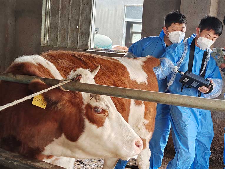
Before performing the scan, the ultrasound probe was cleaned and sterilized to prevent cross infection. The heifer needs to be preferably well bonded before the test so as not to affect the testing process and to ensure the safety and effectiveness of the operation. Generally, if ultrasound machine for cattle testing is also required, the cow must be cleared in advance. First, make sure that the S1 rectal probe is coated with an appropriate amount of ultrasound conductive lubricant. Apply the lubricant evenly to the probe to minimize friction during insertion and to improve smoothness of operation. Next, insert the probe slowly and gently into the cow’s rectum.
During insertion, the operator should proceed with care to ensure that no discomfort or injury is caused to the cow. The depth of insertion usually needs to reach the uterine position so that a systematic examination of the reproductive tract can begin.
Below are some suggested steps and descriptions:
Examine the ovaries:
Begin examining the ovaries, starting from the ovaries to the tip of the uterine horn.
Continue along the uterine horn, traversing the entire horn to reach the body of the uterus.
Follow the curve of the uterus:
Very carefully follow all the curves of the uterus, especially at the tips of the horns. Make sure the movement of the probe is smooth and careful.
Examine all parts of the entire uterus, including the body of the uterus and the horns on either side.
Change the angle of the probe:
The angle of the probe on the rectum is not very important. The angle of the probe can be adjusted in a vertical, horizontal or inclined direction to ensure that the entire area is scanned.
Scanning in longitudinal, transverse or tilted sections:
Scanning can be performed in longitudinal, transverse or tilted sections, which helps to obtain a more comprehensive image.
Adjusting the settings through the control panel of the ultrasound device allows you to adjust parameters such as depth, gain, and frequency to obtain clear images and view a cross-section of the reproductive system in different directions.
Review Verification:
If the operator is unsure of the diagnosis after one examination, a second examination is recommended to validate the test results.
Overall, the operator should meticulously examine the entire reproductive system to ensure that clear images are obtained and to adjust the position and angle of the probe if needed. Maintain care and patience throughout the process to ensure that the examination of the cow is safe and effective.
Post time: Dec-05-2023