


Veterinary ultrasound technology plays an increasingly important role in modern animal medicine, helping veterinarians non-invasively observe internal organs and diagnose diseases in animals. When choosing veterinary ultrasound equipment, we often encounter two types: “color” and “non-color,” which can be confusing for many. So, what exactly is the difference between color and non-color veterinary ultrasound?
I. Imaging Principle and Display Content
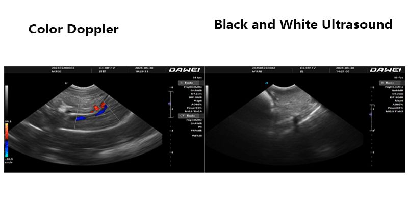
Non-color ultrasound, also known as black-and-white ultrasound or B-mode ultrasound, primarily uses the differences in reflection and scattering of ultrasound waves when they encounter different tissues. These signals are then converted into two-dimensional grayscale images. In the image, fluid usually appears black (anechoic), bones and dense tissues appear white (hyperechoic), and other tissues show varying degrees of gray depending on their density and structure. It mainly displays structural information such as the morphology, size, outline of tissues, and the presence of abnormal masses.
Color ultrasound typically refers to color Doppler ultrasound. It adds the Doppler effect on the basis of B-mode ultrasound. The Doppler effect is the change in frequency of a sound wave when it encounters a moving object. By detecting this frequency change, color Doppler ultrasound can display the direction and speed of blood flow. In a color Doppler image, blood flowing towards the probe usually appears red, and blood flowing away from the probe appears blue, with brighter colors indicating faster flow. In addition, it can also display the nature of blood flow (e.g., laminar flow, turbulent flow), and even the movement information of some tissues themselves.
II. Diagnostic Capabilities and Application Scope
Non-color ultrasound has a wide range of applications in veterinary clinical practice, such as:
- Organ morphological examination: Assessing the size, shape, and echogenicity of internal organs such as the liver, kidneys, bladder, uterus, and spleen, as well as the presence of stones, cysts, tumors, etc.
- Pregnancy diagnosis: Detecting pregnancy in animals and evaluating fetal number, development, and viability.
- Diagnosis of effusions: Detecting pleural effusion, abdominal effusion, pericardial effusion, etc.
- Guidance for puncture: Performing biopsies, fluid aspirations, and other procedures under ultrasound guidance to improve accuracy.
Color ultrasound, due to the added blood flow information, significantly enhances its diagnostic capabilities, especially with unique advantages in the following areas:
- Diagnosis of cardiovascular system diseases: This is the most important application area of color Doppler ultrasound. It can evaluate abnormal heart structures (e.g., valvular disease, cardiac chamber dilation), hemodynamic changes (e.g., regurgitation, stenosis, shunts), and accurately measure blood flow velocity and pressure gradients, which is crucial for diagnosing congenital heart disease, cardiomyopathy, valvular disease, etc.
- Tumor blood supply assessment: Malignant tumors usually have rich blood supply, and color Doppler can display blood flow signals inside and around the tumor, which helps in determining the nature and malignancy of the tumor.
- Inflammation and infection: Blood flow in inflamed areas usually increases, and color Doppler can help identify inflammatory lesions.
- Diagnosis of vascular diseases: Evaluating vascular stenosis, occlusion, thrombosis, etc.
- Assessment of tissue perfusion: Understanding the blood supply to tissues is helpful for the diagnosis and prognosis of certain diseases.
III. Equipment Cost and Operation Difficulty
Generally speaking, color ultrasound equipment is significantly more expensive than non-color ultrasound equipment due to its more complex technology and higher internal hardware and software requirements.
In terms of operation, non-color ultrasound is relatively intuitive, focusing mainly on the grayscale characteristics of the image. Color ultrasound, however, requires operators to have a deeper understanding of hemodynamics and to be proficient in adjusting Doppler modes (such as PRF, gain, filtering, etc.) to obtain high-quality blood flow images, which also increases the difficulty of diagnosis.
IV. Summary and Selection Recommendations
| Feature | Non-Color Ultrasound (Black-and-White Ultrasound) | Color Ultrasound (Color Doppler Ultrasound) |
| Main Function | Displays tissue morphology, size, structure, lesions | Displays tissue morphology, size, structure, and blood flow direction, velocity |
| Advantages | Economical, relatively simple to operate, sufficient for basic diagnosis | Provides blood flow information, strong diagnostic ability for cardiovascular and vascular lesions |
| Disadvantages | Cannot assess blood flow, limited for certain disease diagnoses | High cost, more complex operation, longer learning curve |
| Applications | Basic examinations, pregnancy diagnosis, routine organ assessment, guided puncture, etc. | Heart disease, vascular lesions, tumor blood supply assessment, inflammation, etc. |
When choosing veterinary ultrasound equipment, veterinarians should comprehensively consider their clinical needs, budget, and technical proficiency. If the primary focus is routine organ morphological examinations, pregnancy diagnosis, etc., non-color ultrasound can meet most needs and is economical and practical. However, if in-depth diagnosis of cardiovascular system diseases or complex tumors is required, or if the goal is to improve diagnostic accuracy, then investing in a color ultrasound equipment will be very necessary. Of course, in addition to whether it has color, factors such as the type of probes, image resolution, portability, and after-sales service are also important considerations when purchasing ultrasound equipment.
Post time: Jul-24-2025