


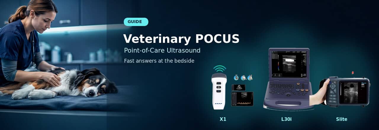
 Point-of-care ultrasound — better known as POCUS — is one of the fastest-growing clinical trends in veterinary medicine. At Dawei Veterinary Medical, we design portable and wireless ultrasound systems that make bedside diagnostics faster and more accessible for small animal practices. In this guide, we explain what veterinary POCUS is, walk through the key protocols, and help you choose the right equipment for your clinic.
What Is Veterinary POCUS?
POCUS stands for Point-of-Care Ultrasound. It refers to focused, real-time ultrasound examinations performed by the attending veterinarian directly at the patient’s location — whether in the consultation room, emergency ward, or beside the cage.
Unlike a complete abdominal ultrasound performed by a radiology specialist, POCUS is designed to answer specific clinical questions quickly:
- Is there free fluid in the abdomen?
- Is there a pericardial effusion?
- Is the bladder intact?
- Is there evidence of pneumothorax?
POCUS is not a replacement for a full diagnostic ultrasound or radiographs. Rather, it is an extension of the physical examination — a rapid bedside tool that helps veterinarians make faster, better-informed decisions in time-critical situations.
To understand the underlying imaging principles behind POCUS, you can read our overview of veterinary ultrasound imaging principles.
Why POCUS Matters in Small Animal Practice
The shift toward POCUS in veterinary medicine mirrors what has already happened in human emergency medicine. Several factors are driving the trend:
- Speed of decision-making — In emergency cases, POCUS can identify life-threatening conditions (hemoabdomen, pericardial effusion, pneumothorax) within minutes of patient arrival.
- Accessibility — Modern portable and wireless ultrasound devices have become smaller, more affordable, and easier to operate, putting real-time imaging into the hands of general practitioners.
- Repeatable and non-invasive — POCUS uses no ionizing radiation and can be repeated as often as needed, making it ideal for monitoring critically ill patients overnight.
- Complements — not replaces — other modalities — POCUS is used as a first-line triage tool. Once the patient is stabilized, full imaging (complete ultrasound, radiographs, or CT) can follow.
Common Veterinary POCUS Protocols
Veterinary POCUS is built around several standardized examination protocols. The most widely used are AFAST, TFAST, and VetFAST.
AFAST (Abdominal Fast Assessment Sonography in Trauma)
AFAST is a rapid abdominal ultrasound protocol designed to detect free peritoneal fluid — typically blood or effusion. The examination uses four standardized views:
- Diaphragmatico-hepatic view (DH) — Evaluates the liver, diaphragm, and gallbladder region for fluid surrounding the liver lobes.
- Spleno-renal view (SR) — Assesses the left kidney and spleen for fluid in the retroperitoneal and peritoneal spaces.
- Cysto-colic view (CC) — Checks the cranial bladder and caudal abdomen for free fluid near the bladder.
- Hepato-renal view (HR) — Examines the right kidney and gallbladder region.
AFAST is especially valuable in trauma cases, post-surgical monitoring, and any patient presenting with a distended abdomen or unexplained collapse. For a deeper look at what abdominal ultrasound reveals in dogs, see our article on what an abdominal ultrasound will show in dogs.
TFAST (Thoracic Fast Assessment Sonography in Trauma)
TFAST focuses on the thorax and is designed to detect:
- Pneumothorax — Using the “lung point” and “gliding sign” to identify absent lung sliding.
- Pleural effusion — Fluid in the pleural space around the lungs.
- Pericardial effusion — Fluid in the pericardial sac, which can cause cardiac tamponade.
TFAST typically uses five probe positions: bilateral perihilar, bilateral chest tube site (CTS), and a subxiphoid (diaphragmatico-hepatic) view for the pericardium.
VetFAST
VetFAST (Veterinary Focused Assessment with Sonography for Trauma) is a broader veterinary adaptation that combines elements of both AFAST and TFAST, and may also include focused cardiac evaluation. Some protocols extend to assess volume status (e.g., inferior vena cava diameter changes) and lung B-lines for pulmonary edema.
Common Clinical Applications of POCUS
Beyond trauma, POCUS is increasingly used in everyday small animal practice for:
| Clinical Scenario | What POCUS Looks For | Key Views |
| Blunt trauma / hit by car | Hemoabdomen, pneumothorax | AFAST + TFAST |
| Acute collapse | Pericardial effusion, cardiac tamponade | TFAST pericardial view |
| Abdominal distension | Ascites, organomegaly | AFAST DH + SR views |
| Dyspnea / respiratory distress | Pleural effusion, pulmonary edema | TFAST CTS bilateral |
| Post-operative monitoring | Internal bleeding at surgical site | AFAST serial scans |
| Rapid pregnancy confirmation | Gestational sac presence | Abdominal uterine scan |
For routine pregnancy ultrasound in dogs, see our complete guide on how to ultrasound a dog for pregnancy.
What Equipment Do You Need for Veterinary POCUS?
POCUS does not require a high-end cart-based ultrasound system. The key equipment requirements are:
- Portability — The device must be easy to carry to the patient, not the other way around.
- Fast startup time — In emergencies, every second counts. The device should power on and be ready in under 30 seconds.
- Appropriate probe — A microconvex probe is the most versatile choice for POCUS. Its small footprint fits into intercostal spaces and provides enough depth for both abdominal and thoracic scanning.
- Image quality sufficient for fluid detection — POCUS primarily looks for anechoic (black) fluid pockets and lung patterns, which do not require the highest-resolution imaging.
- Durability and ease of cleaning — Bedside use means exposure to fur, blood, and fluids. The probe and device should be easy to disinfect.
Dawei portable ultrasound systems are built for exactly these demands — compact, quick to boot, and compatible with microconvex probes suited for POCUS examinations.
If your clinic is looking for a dedicated POCUS device, a wireless ultrasound probe for POCUS that connects directly to a smartphone or tablet can be an excellent entry point — lightweight, always within reach, and ideal for cage-side exams.
Wireless Probe vs Handheld Ultrasound for POCUS
Both wireless ultrasound probes and handheld ultrasound machines are suitable for POCUS, but they serve slightly different needs:
| Feature | Wireless Ultrasound Probe | Handheld Ultrasound Machine |
| Form factor | Probe-only, connects to phone/tablet | All-in-one device with built-in screen |
| Portability | Excellent — fits in a pocket | Very good — small enough to carry room-to-room |
| Battery life | Typically 2-4 hours continuous | Typically 3-5 hours continuous |
| Screen size | Phone or tablet (flexible) | Built-in screen (5-7 inches typical) |
| Best for | Quick cage-side checks, multi-device flexibility | Standalone exams, emergency triage |
| Image review | View on phone/tablet, easy to capture screenshots | Direct on-device, can export via WiFi |
For practices that already use tablets or smartphones in clinical workflow, a wireless probe offers maximum flexibility. For veterinarians who prefer a self-contained device with no phone dependency, a handheld veterinary ultrasound machine provides a dedicated screen and all-in-one operation.
When POCUS Is Not Enough
POCUS is a triage and screening tool — not a comprehensive diagnostic exam. Knowing when to escalate is just as important as knowing how to scan.
Refer for a full ultrasound or advanced imaging when:
- · POCUS identifies an abnormality (e.g., mass, organ enlargement) that requires detailed characterization
- · The patient is stable enough for a thorough examination and the clinical question goes beyond “is there fluid?”
- · Cardiac evaluation requires Doppler assessment of valvular flow and chamber measurements
- · Radiographs or CT are needed for skeletal, thoracic wall, or detailed abdominal evaluation
For cases requiring vascular flow assessment, color flow mapping, or spectral Doppler analysis, a veterinary Doppler ultrasound for small animals provides the advanced imaging capabilities that POCUS devices cannot offer. To understand the difference between imaging modes, see our comparison of B-mode vs Doppler ultrasound.
Choosing a Dawei Ultrasound Solution for POCUS
Dawei Veterinary Medical offers several ultrasound platforms suitable for point-of-care use in small animal practice. Here is a quick guide to matching the right system to your clinical needs:
| Clinical Need | Recommended Dawei Solution | Why |
| Cage-side POCUS in emergency ward | Wireless ultrasound probe system for small animals | Pocket-sized, instant connection to phone/tablet |
| General practice POCUS + routine exams | Handheld veterinary ultrasound diagnostic machine | Dedicated screen, versatile probe options |
| Full abdominal and cardiac exams with Doppler | Pet ultrasound machine with veterinary Doppler system | Color/PW Doppler for comprehensive diagnostics |
| Multi-room portable use | Portable wireless ultrasound category | Flexibility across multiple examination rooms |
To explore the full range of portable wireless ultrasound options, browse our wireless ultrasound collection.
FAQ About Veterinary POCUS
What does POCUS mean in veterinary medicine?
POCUS stands for Point-of-Care Ultrasound. It is a focused, bedside ultrasound examination performed by the attending veterinarian to answer specific clinical questions in real time — such as detecting free abdominal fluid, pericardial effusion, or pneumothorax.
Is POCUS the same as a full abdominal ultrasound?
No. POCUS is a rapid, targeted examination (typically 5-10 minutes) designed to detect specific findings like free fluid or lung pathology. A full abdominal ultrasound is a comprehensive, systematic examination of all abdominal organs performed by a trained specialist, typically taking 30-45 minutes.
What probe is best for veterinary POCUS?
A microconvex probe is the most commonly recommended probe for veterinary POCUS. Its small footprint fits well in intercostal spaces for thoracic scanning, and it provides sufficient depth and field of view for abdominal views. Frequency range of 5-8 MHz is typical for small animal POCUS.
Can POCUS be used in emergency veterinary care?
Yes. POCUS originated in human trauma medicine and has become a standard triage tool in veterinary emergency care. AFAST and TFAST protocols are specifically designed for rapid assessment of trauma patients and can identify life-threatening conditions within minutes of presentation.
Is wireless ultrasound suitable for POCUS?
Yes. Wireless ultrasound probes are well-suited for POCUS because they are lightweight, connect directly to a smartphone or tablet, and can be carried in a pocket. They are ideal for quick cage-side assessments and emergency triage where a full ultrasound machine may not be immediately available.
When should a veterinarian choose Doppler ultrasound instead of basic B-mode?
Doppler ultrasound is necessary when the clinical question involves blood flow — such as assessing cardiac valvular regurgitation, measuring vascular flow velocity, or evaluating organ perfusion. For basic POCUS applications (fluid detection, lung sliding, organ size), B-mode imaging is sufficient. Learn more in our guide to B-mode vs Doppler ultrasound.
Conclusion
Veterinary POCUS is transforming how small animal practices approach triage, emergency care, and daily clinical decision-making. With the right portable equipment, any veterinarian can perform rapid, focused ultrasound exams that save critical minutes when they matter most.
Dawei Veterinary Medical is committed to making point-of-care imaging accessible, affordable, and practical for clinics of all sizes. Whether you need a wireless probe for cage-side checks or a handheld system for comprehensive portable diagnostics, our ultrasound platforms are designed to support your POCUS workflow.
Ready to equip your practice for POCUS? Contact our team to find the right ultrasound solution for your clinical needs.
Post time: Jul-27-2026