


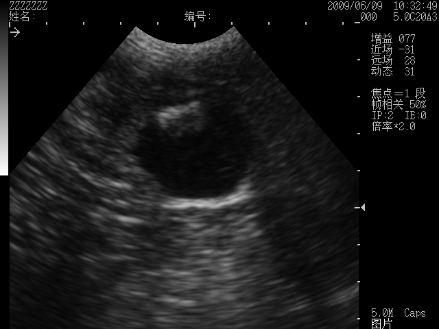
1. Bladder stones During a bladder ultrasound examination of a dog using a dog ultrasound machine, the standing bailout probe is swept upward over the bladder of the dog, in which case the free stone appears above the bladder image and carries an acoustic shadow. The image obtained with the standing bowing probe from the bottom up. The liquid dark area is the bladder, and the strong echoes in the upper part of this area are the images of the stones.
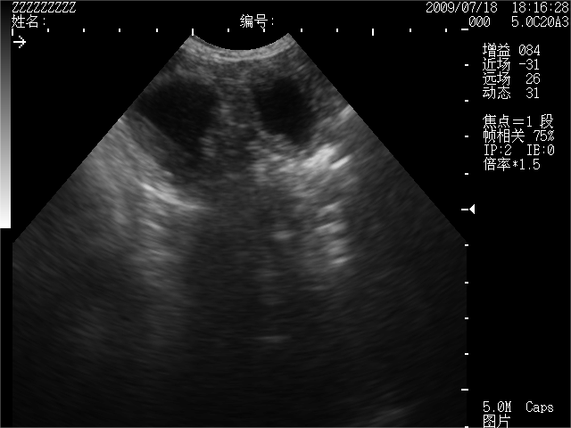
2. Uterine pus accumulation During uterine exploration, a transverse view of the uterine horns is taken and the canine uterine pus is visualized. The image shows a transverse section of the uterine horn junction, and the two fluid dark areas in the image show fluid accumulation within the uterine horn.
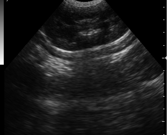
3. Kidney stones Imaging image of a canine kidney stone. The strongly echogenic light mass in the middle of the kidney in this image is a stone.

4. Prosta cyst image. This image shows fluid that has started in the prostate. Abminal ultrasound using a dog ultrasound machine can help provide accurate and safe diagnostic information.Ultrasound clearly shows a dog’s internal organs, including the heart, liver, kidneys, bladder, and more, to help veterinarians assess their health and function.
Abminal ultrasound using a dog ultrasound machine can help provide accurate and safe diagnostic information.Ultrasound clearly shows a dog’s internal organs, including the heart, liver, kidneys, bladder, and more, to help veterinarians assess their health and function.
Post time: Apr-16-2024