


Indications for uterine ultrasound in dogs
(1) Routine abdominal ultrasound should include the uterus;
(2) Sterilized animals should be routinely examined for uterine body segments and ovarian segments;
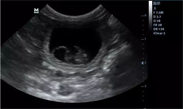
(3) Pregnancy diagnosis;
(4) Vaginal discharge;
(5) Hematuria;
(6) Suspicion of uterine infection;
(7) Abdominal mass suspected of originating in the uterus;
(8) Enlargement of the entire abdomen;
(9) Abdominal or pelvic pain;
Methods of finding the uterus in dogs
(1) Locate the body of the uterus by means of the inflated colon and fluid-filled bladder (this is also known as “triangulation”).
(2) Find the ovaries behind both kidneys.
(3) The angle of the uterus between the posterior kidneys and the body of the uterus is variable and is not normally seen by default when the equipment is not clear enough.
Normal ultrasound images of the uterus in dogs
(1) When normal, it is often not detected by scanning. If the technique is correct, it is normal by default if it is not detected by scanning.
(2) The uterus is a homogeneous hypoechoic luminal structure without any layering of the uterine wall during interoestrus and lesions.
(3) A hyperechoic streak may be present in the uterine lumen during pre-estrus, estrus, and early inter-estrus.
(4) Uterine images may vary by breed. For example, medium to large dogs may have a small, uniform area of fluid in the uterus when normal, which is not abnormal.
(5) At 2-3 weeks post-pregnancy, the internal diameter of the uterus is enlarged and there is a productive echogenic material inside, which may be malaise. It is a normal condition.
Dawei Veterinary Medical, as a professional dog ultrasound machine manufacturer in China, has provided veterinary equipment services for more than two hundred countries around the world, if you have any related needs, welcome to come and consult us!
Post time: Mar-21-2024