


1. Microconvex probe advantages for cats cans
In the rapidly evolving landscape of veterinary medicine, diagnostic imaging has become a cornerstone of daily practice for small animal clinics. Among the various tools available to the modern veterinarian, the ultrasound machine stands out as a non-invasive, real-time diagnostic modality that provides critical insights into internal medicine cases. However, the quality of these insights is inextricably linked to the choice of transducer. For mixed-practice veterinarians and small animal specialists alike, the question of “which probe to buy first” often yields a unanimous answer: the microconvex probe.
This article serves as a comprehensive technical guide for veterinary professionals seeking to optimize their imaging workflow for feline and canine patients. We will explore the physics behind the microconvex probe’s design, its specific advantages over linear and phased array alternatives, and why it remains the “workhorse” of the veterinary clinic. Whether you are performing a routine abdominal scan on a geriatric cat or a focused assessment with sonography for trauma (FAST) on a large breed dog, understanding the nuances of your transducer is the first step toward diagnostic confidence. We will also examine the practical applications of this technology, supported by authoritative clinical evidence and a detailed look at modern equipment like the DAWEI Veterinary L30i. By the end of this deep dive, you will have a clear understanding of why “Microconvex probeadvantages for cats cans” is not just a search term, but a fundamental truth of veterinary imaging.

1.1 What is a Microconvex Probe in Veterinary Ultrasound?
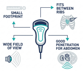
To appreciate the utility of the microconvex probe, one must first understand its engineering and physical characteristics. A microconvex probe, often referred to as a “micro-curved” or “small radius” array, is a transducer designed specifically to bridge the gap between the deep penetration of a standard convex probe and the high resolution of a linear probe. Unlike the large curvilinear probes used in human medicine for abdominal scanning—which often have a footprint too large for small animal patients—the microconvex probe features a significantly reduced radius of curvature. This compact design allows the piezoelectric crystals to fan out the ultrasound beam from a very small point of contact, creating a wide, pie-shaped sector image that expands as it travels deeper into the tissue.
The “micro” designation refers primarily to this small footprint, which is the defining feature that makes it indispensable for veterinary use. In clinical practice, the footprint is small enough to fit comfortably between the ribs of a medium-sized dog or tucked under the costal arch of a small cat, yet the curvilinear array provides a wide field of view in the far field. This geometry is crucial because it allows the operator to visualize large organs like the liver or widespread pathologies like ascites through a tiny acoustic window.
Veterinary ultrasound machines equipped with a microconvex probe typically operate in a frequency range of 5.0 MHz to 8.0 MHz, though modern broad-bandwidth probes can often span from 3.0 MHz up to 9.0 MHz. This frequency range strikes a critical balance: it is high enough to provide excellent spatial resolution for distinguishing the layers of the feline intestine or the architecture of the canine kidney, yet low enough to penetrate to the depth required for a thorough abdominal survey in all but the largest giant-breed dogs.
For the general practitioner, the microconvex probe is the “desert island” transducer—the one probe you would choose if you could only have one. It is the primary tool for:
- Complete Abdominal Surveys: Scanning the entire abdomen from the liver and biliary tract cranially to the urinary bladder and prostate caudally.
- Pregnancy Diagnosis: Confirming viability and estimating gestational age in both cats and dogs from as early as 21-25 days.
- Emergency FAST/POCUS: Rapidly assessing trauma patients for free fluid (hemoabdomen) or pericardial effusion in a triage setting.
- Thoracic Windows: Viewing the heart and pleural space through intercostal and sub-xiphoid approaches where larger probes cannot maintain contact.
- Guided Procedures: Assisting in cystocentesis or fine-needle aspiration (FNA) of abdominal masses due to the wide visualization field.
1.2 Microconvex Probe Advantages for Cats and Dogs
1.2.1 The Small Footprint Advantage
The most immediate benefit is the physical size of the probe’s face. In cats and small dogs, the space between the last rib and the iliac crest—the flank—is limited. A standard linear probe, which has a long, flat footprint, often cannot make full contact with the curved, concave surface of a thin cat’s abdomen. Air gaps between the probe and skin result in “dropout” artifacts where no image is generated. The small, curved surface of the microconvex probe presses easily into these soft tissue spaces, ensuring 100% coupling efficiency and eliminating air artifacts. As noted by experts in veterinary imaging:
1.2.2. Intercostal Access for Hepatic Imaging
The liver is located well within the rib cage in most dogs and cats. To visualize the hepatic parenchyma, gall bladder, and porta hepatis, the veterinarian must scan between the ribs (intercostal) or angled steeply under the xiphoid process (subcostal). A wide linear probe bridges across the ribs, causing the bone to cast dark acoustic shadows that obscure the liver. The microconvex probe fits snugly between the ribs, utilizing the intercostal muscles as an acoustic window to see the liver clearly without rib shadowing interference.
1.2.3. The “Sector” Field of View
Despite its small contact point, the microconvex probe generates a sector-shaped image that widens with depth. This is particularly advantageous when scanning organs that are larger than the acoustic window itself. For example, a dog’s full urinary bladder may be 5-8 cm in diameter, but the probe contact area is only 2 cm. A linear probe would only show a vertical slice of the bladder equal to its own width. The microconvex probe, however, fans the beam out, allowing the clinician to visualize the entire bladder, including the apex and trigone, from a single central point of contact.
1.2.4. Feline-Specific Ergonomics
Cats are notoriously sensitive patients. High-stress handling can elevate cortisol levels and alter physiological parameters like heart rate and kidney blood flow. The microconvex probe allows for “gentle” scanning. Because it requires a smaller area of clipped fur and less pressure to maintain contact, examinations can often be performed with the cat in a natural sternal position or resting comfortably on a blanket, rather than being forcefully restrained in dorsal recumbency. This “less is more” approach significantly improves feline compliance.
1.2.5. Depth Versatility for Canine Patients
Dogs range in size from 2kg Chihuahuas to 80kg Mastiffs. A high-frequency linear probe (e.g., 12 MHz) is excellent for the Chihuahua’s superficial gut but useless for the Mastiff’s deep spleen. The microconvex probe covers the middle ground perfectly. By adjusting the frequency setting on the veterinary ultrasound machine (e.g., dropping to 5.0 MHz for large dogs or increasing to 8.0 MHz for small dogs), a single probe can serve 90% of the canine patients entering the clinic, making it the most cost-effective investment for a practice.
1.2.6. Improved Gastric and Intestinal Imaging
While linear probes are superior for detailed layering of the gut wall, microconvex probes are often better for the initial survey of the gastrointestinal tract. Their wide field of view helps in following the path of the duodenum or locating the ileocolic junction, which can be disoriented and tortuous. Once the pathology (e.g., a foreign body or mass) is located with the microconvex probe, the clinician can then switch to a linear probe for a high-resolution detailed look if needed, but the microconvex is the “finder” tool.
1.3.Clinical Applications & Scanning Tips (Cats, Dogs, and Beyond)
Implementing the microconvex probe into your daily clinical workflow requires attention to preparation and technique. Even the most advanced ultrasound machine will yield poor images if the fundamental steps of patient preparation and probe handling are neglected.
Step 1: Patient Preparation Ultrasound waves do not travel through air or hair. For high-quality diagnostic images, the patient must be shaved closely to the skin. In cats, clip from the xiphoid to the pubis, extending laterally to the flanks. In dogs, ensure the clip is wide enough to include the lateral intercostal spaces for liver and spleen access. Apply acoustic coupling gel liberally. For best results, allow the gel to sit on the skin for 1-2 minutes to soften the keratin layer before scanning. Alcohol can be used to wet the skin before gel application, but avoid it if you plan to use electrical leads (ECG) as it can degrade contact quality.
Step 2: Probe Grip and Positioning Hold the microconvex probe near the face (the scanning surface), not at the end of the cable strain relief. This “pencil grip” allows for fine motor control and subtle angulation movements (fanning, rocking, and sliding) which are essential for sweeping through entire organs. Stabilize your hand by resting your pinky finger or wrist gently on the patient’s body; this prevents the probe from sliding off the target when the animal breathes or moves.
Step 3: Frequency and Depth Optimization Always start with the highest frequency setting that still allows you to see the bottom of the abdomen. For a cat, start at 7.5-8.0 MHz. For a Labrador, you may need to drop to 5.0 MHz. Adjust your depth so that the organ of interest fills 75% of the screen. A common mistake is leaving the depth set to 15cm when scanning a 3cm cat kidney, resulting in a tiny image lost in a sea of black space.
Step 4: Systematic Exam Protocol Consistency is key to avoiding missed diagnoses. Develop a routine loop, such as:
- Bladder & Prostate/Uterus: Start caudally where the bladder is easy to find. Evaluate wall thickness and luminal content.
- Left Kidney & Spleen: Move cranially along the left flank. Compare the cortical echogenicity of the kidney to the spleen.
- Liver & Gall Bladder: Angle under the rib cage near the xiphoid. Asses for masses, biliary sludge, or hepatic lipidosis (common in cats).
- Right Kidney & Duodenum: The right kidney is more cranial, often tucked under the ribs (fossa for the right kidney). The microconvex probe is essential here to look “up” under the ribs.
- Mid-Abdomen: Sweep the mesentery for lymph nodes and scan the intestinal loops.
| Pro Tip: Microconvex vs. Linear
While the microconvex is the versatile master, the linear probe has its place. Use a Linear Probe when: checking superficial structures like the feline intestinal wall layers (to distinguish IBD from lymphoma), examining the thyroid glands, or looking at superficial eyes/tendons. Use a Microconvex Probe when: you need depth (large dogs), wide field of view (global organ assessment), or access between ribs (liver/spleen). |
1.4 Microconvex for Veterinary POCUS & Lung Ultrasound
One of the most rapidly growing areas in veterinary emergency medicine is Point-of-Care Ultrasound (POCUS), particularly for thoracic and lung applications (Vet BLUE protocols). In patients with respiratory distress (dyspnea), minimizing stress is paramount. You cannot restrain a dyspneic cat for radiographs without risking decompensation. The microconvex probe allows for a rapid, low-stress “flash” scan of the chest while the patient remains standing or sternal.
When scanning the lungs, the goal is to identify artifacts like “B-lines” (or ultrasound lung rockets), which indicate fluid in the interstitium (pulmonary edema, contusions), or the absence of “Glide Signs,” which suggests pneumothorax. The microconvex probe is uniquely suited for this because the lung surface is best viewed through the intercostal spaces. A large convex probe would bridge the ribs, blocking the view of the pleura. A linear probe can work but often has a long footprint that doesn’t sit flat against the curved chest wall of a small dog.
The concave shape of the microconvex face matches the curvature of the intercostal space, allowing the ultrasound beam to bypass the ribs entirely and focus on the pleural line. This creates a clear window to assess for “wet” vs. “dry” lungs. Recent reviews in veterinary literature affirm this utility:
For a “T-FAST” (Thoracic FAST) exam, the veterinarian places the microconvex probe at specific points (Chest Tube Site, Pericardial Site, Diaphragmatico-Hepatic View) to rule out pneumothorax, pericardial effusion, and pleural effusion within minutes of the patient’s arrival.
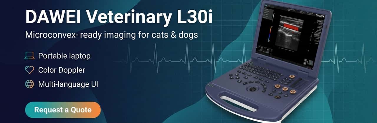
4.Product Spotlight: DAWEI Veterinary L30i (Portable Color Doppler)
For veterinary clinics looking to upgrade their imaging capabilities with a system that maximizes the potential of microconvex technology, the DAWEI Veterinary L30i offers a compelling balance of performance, portability, and modern features. Designed as a laptop-style portable unit, it is particularly well-suited for mobile vets, mixed practices, and space-constrained clinic rooms.
Crucially, the L30i supports a wide range of probe frequencies. The standard convex probe covers 2.0-5.5MHz, while the system supports high-frequency linear imaging up to 12.0MHz. For the all-important microconvex applications, the system’s beamforming and processing power (including Tissue Harmonic Imaging and Spatial Compounding) ensure that the wide-sector images remain crisp from the near field to the far field.
The system includes robust Color Doppler capabilities (Spectral Pulsed Doppler, Power Doppler), essential for distinguishing vascular structures from biliary ducts or evaluating blood flow in organs. With a 128GB SSD, image storage and retrieval are fast, and the inclusion of DICOM 3.0 means studies can be seamlessly sent to PACS servers or teleradiology
|
Feature |
Specification (Source: DAWEI L30i Specs) | |
| Form Factor | Portable Notebook / Laptop Style | |
| Display | ≥15-inch High-Definition Monitor | |
| Imaging Modes | B, 2B, 4B, M, Color Doppler (CFM), Power Doppler (PDI), PW Doppler | |
| Image Tech | Spatial Compounding, Tissue Harmonic Imaging, Trapezoidal Imaging | |
| Storage | ≥128GB SSD; BMP/JPEG/DCM formats; Cine Loop ≥600 frames | |
| Connectivity | 6 USB ports, HDMI, RJ-45 (Network), DICOM 3.0 | |
| Language Support | Chinese, English, French, Russian, Spanish | |
| Probe Options |
Convex (2.0-5.5MHz), Linear (6.0-12.0MHz), Microconvex/Cavity (4.5-9.0MHz) |
|
Clinics seeking a versatile, cost-effective solution for small animal imaging should consider evaluating the L30i for their diagnostic suite.
FAQ: Microconvex Probe & Small Animal Ultrasound
Q1: Is a microconvex probe good for cats? Absolutely. It is arguably the best all-around probe for feline patients. Its small face fits easily under a cat’s costal arch and between ribs, and its frequency range (typically 5-8 MHz or higher) provides excellent resolution for the small organs of a cat.
Q2: Microconvex vs linear probe for cat abdomen: Which is better? For a general overview (“big picture”), the microconvex is better because it shows entire organs in one view. For detailed analysis of the intestinal wall layers (e.g., measuring wall thickness for IBD) or looking at superficial structures, the linear probe is superior due to higher frequency and better near-field resolution. Ideally, you use the microconvex to find the lesion and the linear to measure it.
Q3: What probe is best for dog pregnancy ultrasound? The microconvex probe is the gold standard for canine pregnancy diagnosis. Its wide sector view allows you to count fetuses more accurately and scan the entire uterine horn. It also penetrates deep enough to see fetuses in large dogs, whereas a linear probe might not reach deep enough.
Q4: Can microconvex be used for FAST/POCUS exams? Yes, it is the preferred probe for A-FAST (Abdominal) and T-FAST (Thoracic) exams. Its small footprint allows for quick placement at the four standard acoustic windows without needing to shave large areas of the patient in an emergency.
Q5: What frequency range is typical for a veterinary microconvex probe? Most microconvex probes operate between 4.0 MHz and 9.0 MHz. Lower frequencies (4-5 MHz) are used for large dogs, while higher frequencies (7-9 MHz) are used for cats and small dogs.
Q6: How to choose a veterinary ultrasound machine for a small animal clinic? Look for a system that supports a wide range of probes (must have microconvex and linear ports), has “veterinary-specific” presets (not just human presets renamed), and offers DICOM connectivity for telemedicine. Portability is also key if you move between consult rooms or exam tables.
Conclusion
In conclusion, the microconvex probe is the unsung hero of the veterinary ultrasound world. Its unique combination of a small footprint, wide field of view, and versatile frequency range makes it the single most useful transducer for mixed small animal practice. It solves the anatomical challenges presented by cats and dogs—namely, small acoustic windows and variable body sizes—better than any other probe type. Whether you are investing in your first system like the DAWEI Veterinary L30i or upgrading your current setup, ensuring you have a high-quality microconvex probe is the best way to guarantee diagnostic success. By mastering this tool, veterinarians can perform faster, more accurate, and less stressful exams, ultimately leading to better patient outcomes.
Post time: Feb-11-2026