


L3-VET Portable laptop veterinary ultrasound scanner can be used for the examination of different organs in sheep, dogs, cats, reptiles, etc. The images are preset to optimize the examination conditions and reduce the adjustment during veterinary operation.
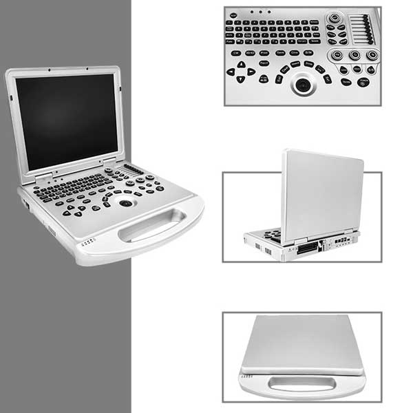
L3-VET Portable laptop veterinary ultrasound
I. Measurements
1. Routine Measurements
All operations must be performed in frozen state.
B-mode routine measurements include: Distance, Vessel Stenosis Rate, Angle, Circumference/Area, Area Stenosis Rate, Volume, etc.
Steps:
- Use the trackball to move the cursor to the desired measurement item and press the [Set] key.
- Move the cursor to the measurement start point and press [Set].
- Move the cursor to the measurement end point and press [Set].
- Measurement results will display at the bottom of the screen.
- Press [Clear] to delete the measurement.
- Press [Freeze] to exit the measurement.
2. Heart Rate Measurement
The system calculates heart rate by averaging multiple cardiac cycles for improved accuracy.
In B/M or M Mode:
- In frozen state, move the cursor to “M Routine” in the measurement menu and press [Set].
- Select “Heart Rate” and press [Set].
- Mark the start point (beginning of two cardiac cycles) with [Set].
- Mark the end point (end of two cardiac cycles) with [Set].
- Repeat steps 3–4 for multiple measurements.
- [Clear]: Delete data.
- [Freeze]: Exit.
Note: For higher accuracy, perform multiple measurements and average the results.
PW Doppler Submenu:
- Time, Heart Rate, Velocity, Acceleration, Doppler Routine, Doppler Trace.
-
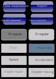
Heart Rate in D Mode:
- In frozen D mode, select “Routine Measurement” > “D Routine” > “Heart Rate”.
- Mark start and end points of two cardiac cycles.
- Results display at the lower right of the screen.
3. Velocity Measurement (Doppler Mode)
Measures velocity and pressure gradient at a specific point on the Doppler spectrum.
Steps:
- In frozen D mode, select “Routine Measurement” > “D Routine” > “Velocity”.
- Mark start and end points on the waveform.
- Results display at the lower right.
4. Obstetric Measurements
Used to estimate gestational age and due date using species-specific obstetric tables.
Steps:
- In B mode, freeze the image and select “Early/Mid-Late Gestation” from the measurement menu.
- Choose a measurement item (e.g., GS: Gestational Sac):
- Mark start and end points with [Set].
- Repeat for multiple planes (e.g., measure GS 3 times for averaging).
- Other parameters (CRL, BD, HD, BPD, TD) follow similar steps.
Key Parameters:
- GS: Gestational Sac
- CRL: Crown-Rump Length (body length)
- BD: Body Diameter
- HD: Head Diameter
- BPD: Biparietal Diameter
- TD: Trunk Diameter
Note:
- Measurements are based on formulas from Veterinary Imaging; results may vary by species.
- Partial data is retained after unfreezing.
II. Saving and Generating Reports
- In frozen state, press the [Report] key.
- Select a report template.
- On the report interface:
- Use “Diagnostic Template” (lower right) to load predefined templates for specific anatomical regions.
- Fill in details and select “Print Preview” to review.
- Options:
- “Print”: Directly print the report.
- “Close”: Exit without saving.
- “Return”: Go back to the previous screen.
Operational Essentials:
- Master three core measurements: Routine, Heart Rate, and Obstetric.
- Always freeze the image before measuring.
- For obstetrics, average multiple scans for accuracy.
Post time: May-15-2025