


In modern dairy farming, achieving “accurate, timely, and correct breeding” has become the key to improving reproductive efficiency. As herd sizes continue to grow, traditional methods that rely on visual observation or behavioral signs to detect estrus and ovulation are often inaccurate and inefficient. Bovine ultrasound for monitoring dairy cow follicle development is increasingly becoming a standard practice in large farms, breeding enterprises, and reproductive service teams.
Why Use Ultrasound to Monitor Follicles?
Estrus signs in dairy cows can vary greatly. High-yield cows often show silent estrus, making it difficult for staff to accurately determine the reproductive stage. Although follicles are internal, veterinary ultrasound can clearly visualize them as round or oval low-echo structures, allowing direct observation of diameter, wall thickness, and arrangement.
This enables reproduction technicians to rely on objective imaging data rather than experience alone.
Follicle development follows physiological patterns:
-
When a follicle reaches 10–16mm, it usually enters the dominant follicle stage.
-
When it approaches 18mm or more, it is often close to ovulation.
With devices such as Dawei waterproof veterinary bovine ultrasound machines, follicle diameters can be measured with high resolution, and boundaries can be marked for more accurate assessment.
Interpreting Reproductive Status from Follicle Imaging
When examining a cow’s ovaries with a dairy cow ultrasound machine, technicians typically focus on:
-
The presence of multiple follicle-like structures on the ovarian surface, indicating active follicular waves.
-
Dominant follicles, which usually appear with uniform echogenicity and clear contours.
-
Regressing follicles, which present irregular shapes and uneven echoes.
-
Pre-ovulatory follicles, which show thinning walls and increased tension in the image.
Experienced technicians can estimate if a cow is in the 12-hour window before ovulation, providing a more reliable basis for reproductive management than traditional visual observation. This is particularly important for high-yield cows with subtle estrus behavior, where follicle imaging is often the only reliable way to determine the optimal artificial insemination timing.
Determining Breeding Timing: The Value of Ultrasound Imaging
Follicle development is a dynamic process. A veterinary ultrasound machine for cows can accurately show:
-
Whether the follicle has entered the dominant stage
-
Whether ovulation is approaching
-
Presence of a corpus luteum (post-ovulation marker)
-
Ovarian inactivity or cyst formation
In practice, technicians often determine the AI timing by measuring follicle size. When a follicle reaches 18–20mm with a thinning wall, ovulation usually occurs within 6–18 hours, making this the ideal time for artificial insemination to maximize conception rates.
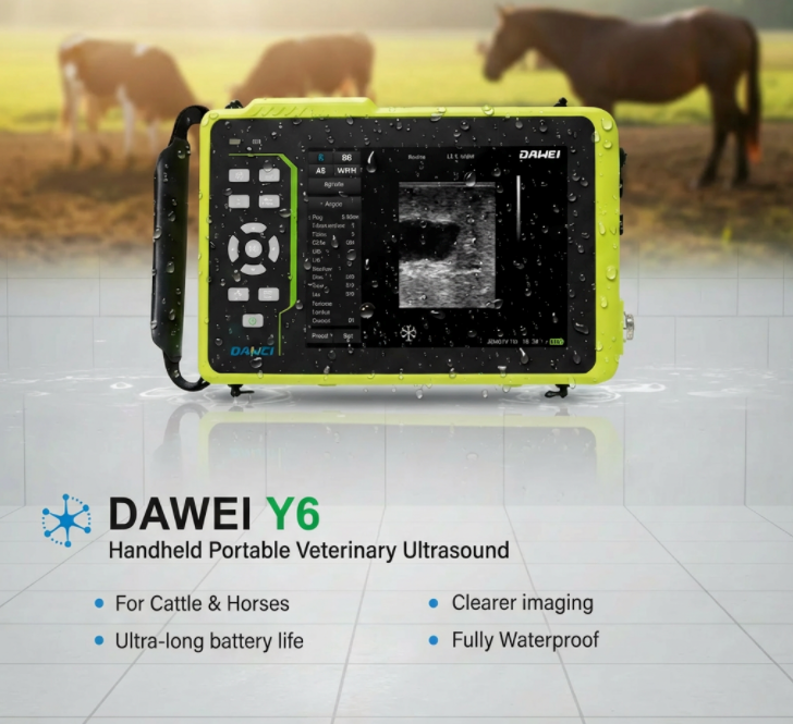
For less experienced staff, using devices like the Dawei Y6 veterinary bovine ultrasound allows direct measurement of follicle size. The system can outline follicle boundaries in real-time and provide millimeter-level diameter readings, reducing human error and improving overall reproductive success.
Conclusion
Monitoring dairy cow follicle development with a bovine ultrasound machine has become an essential tool in modern dairy farms. It makes reproduction management more scientific, significantly improves conception rates, reduces days open, and helps farms maintain a stable production rhythm.
Post time: Dec-25-2025