


During the periparturient period (three weeks before to three weeks after calving), feeding and management are major challenges. If your cow shows poor appetite, reduced milk yield, or even ketosis after calving, she may be suffering from a hidden metabolic disorder—fatty liver syndrome.
Fatty liver is a major health threat for dairy cows in the periparturient period and significantly affects both health and economic performance. But how can we diagnose it quickly and accurately?
Liver biopsy is accurate but invasive, time‑consuming, and risky, making it unsuitable for large‑scale clinical or farm use.
I. Why Is Bovine Ultrasound the First Choice for Diagnosing Fatty Liver?
Ultrasound offers several key advantages:
• Non‑invasive: No blood sampling or tissue collection; almost painless for the cow.
• Fast and easy: The exam takes only a few minutes and results appear instantly.
• High reliability: Studies show that ultrasound has good sensitivity and specificity for detecting hepatic fat infiltration and is a practical alternative to biopsy.
Ultrasound examinations are primarily performed during the periparturient period, the critical window when fatty liver develops.
II. Ultrasound Examination: Where to Scan and How?
To assess the cow’s liver, the ultrasound probe is placed at a specific location:
1. Scanning Site: The right 10th–12th intercostal spaces, caudal to the costal arch and near the lower edge of the scapula.
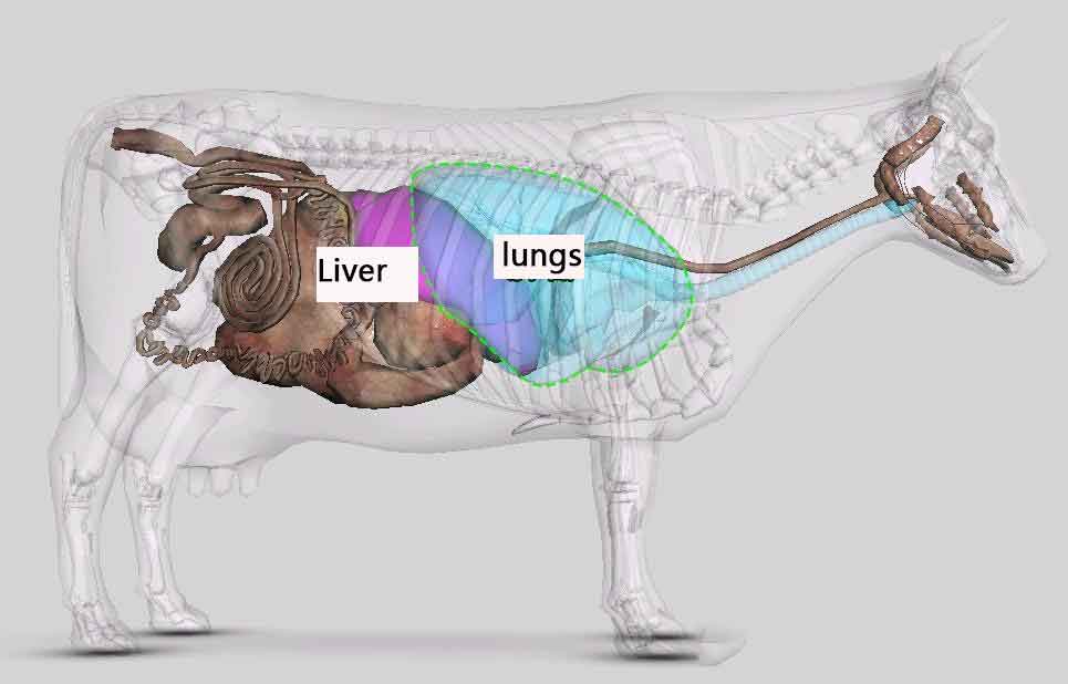
The liver is mainly located in the right cranial abdomen, partially obscured by the diaphragm.
Scanning at the 10th–12th intercostal spaces avoids pulmonary interference and provides clearer hepatic images.
2. Probe Selection: A 3.5 MHz convex or linear probe is commonly used.
3. Procedure: Clip hair, apply coupling gel, and place the probe parallel to the ribs to scan from multiple angles.
Steps:
• Stand on the cow’s right side, observing from behind the shoulder.
• Count to the 10th–12th ribs.
• Apply gel below the right costal arch at the costo‑diaphragmatic region.
• Hold the probe perpendicular or slightly cranially angled 15–30°.
• Move slowly to locate uniform gray‑white liver parenchyma without major pulsating vessels.
III. Four Key Ultrasound Features of Fatty Liver
Fat accumulation changes the acoustic properties of liver tissue. On ultrasound, fatty liver displays distinct characteristics:
1. Increased Echogenicity: The liver appears brighter/whiter than normal due to higher fat reflectivity.
2. Coarser Texture: Parenchymal patterns become uneven and coarse.
3. Blurred Vessels: Vessel margins (portal vein, hepatic vein) become indistinct or invisible.
4. Deep Attenuation: Ultrasound penetration decreases significantly; deeper tissues appear darker because fat absorbs sound waves.
Among these, increased echogenicity and deep attenuation are the most important indicators of fatty liver severity.
IV. Fatty Liver Grading: Three‑Level Diagnostic System
Based on severity of ultrasound features, fatty liver can be divided into four grades to guide treatment and management:
Grade | Clinical Reference | Ultrasound Criteria
Normal | No clinical signs | Uniform echogenicity; clear vessel margins
Grade 1 (Mild) | Mild or no signs | Increased echogenicity with vessel blurring; no marked deep attenuation
Grade 2 (Moderate) | Noticeable signs (e.g., decreased feed intake) | Increased echogenicity, vessel blurring, and obvious deep attenuation
Grade 3 (Severe) | Severe symptoms (e.g., recumbency) | Marked echogenicity; severe attenuation with vessels not visible
Ultrasound grading helps differentiate normal, moderate, and severe fatty liver based on image quality and attenuation level.
Using bovine ultrasound is a highly valuable tool for managing dairy cows during the periparturient period. Farms and veterinarians should make full use of this method to routinely monitor high‑risk cows such as over‑conditioned or high‑yield individuals.
Early detection and grading of fatty liver enable timely diet adjustment, energy supplementation, and medical treatment to reduce metabolic disease risks and protect herd health and productivity.
Post time: Dec-03-2025