


In modern cattle breeding management, accurately identifying whether a cow is pregnant, the stage of pregnancy, and the development of the embryo is critical to improving reproductive efficiency and economic benefits. Traditional rectal palpation relies heavily on experience and is subjective. With the advancement of imaging technology, veterinary ultrasound has become an essential tool for pregnancy diagnosis and determining gestational age in cows.
I. Principle of Pregnancy Detection with Ultrasound
Using a rectal probe, ultrasound waves are transmitted into the cow’s body and reflected to form images, allowing observation of the uterus and ovaries. At different stages of pregnancy, the ultrasound images display distinct features, such as uterine fluid, embryo length, fetal heartbeat, and placental structures. By analyzing these indicators, veterinarians and technicians can scientifically determine the cow’s gestational days.
II. Ultrasound Characteristics at Different Stages
21 days pregnant: No clear embryo sac is visible yet. Color Doppler mode can be used to check corpus luteum blood flow to determine pregnancy.
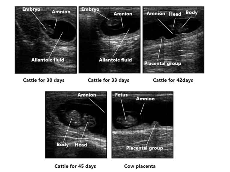
25–30 days pregnant: The embryo becomes visible, and by day 30, the amniotic and allantoic membranes can be seen.
35–45 days pregnant: The embryo is more developed, with visible limbs and head. Fetal heartbeat can be observed, making gestational age estimation more accurate.
Around 60 days pregnant: The fetus has a complete structure. Fetal length can be measured to determine gestational age, and gender determination becomes possible.
Over 90 days pregnant: The fetal skeleton calcifies, image clarity improves, and measurements such as biparietal diameter and femur length can be used to estimate fetal age.
III. Methods for Determining Gestational Age
Embryo length observation: From 30–60 days, embryo length strongly correlates with gestational age.
Specific measurements: After 90 days, biparietal diameter and femur length provide reliable fetal age estimation.
Fetal heartbeat: Combining heart rate with developmental stage increases accuracy.
Reference curve: Farms can establish their own “pregnancy growth curve” for higher precision.
IV. Application Value
Shortening open days: Early detection helps identify non-pregnant cows quickly for rebreeding.
Scientific calving management: Predict calving time and prepare feed and management plans in advance.
Improving conception rates: Avoid repeated insemination and reduce economic loss.
Supporting breeding research: Ultrasound data can be used to study reproductive performance under different breeds and conditions.
Recommended Device:Dawei Y6 Cattle Equine Portable Vet Ultrasound Machine Device
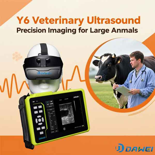
To help cattle farms achieve more efficient reproductive management, Dawei introduces the Y6 Waterproof Veterinary Ultrasound Scanner.
Designed for cattle: Rectal probe ensures accurate pregnancy diagnosis
8-inch HD display: Clear images for embryo sac, heartbeat, and skeletal structures
Fully waterproof body: Durable and reliable for farm environments
Comprehensive measurement functions: Supports fetal length, biparietal diameter, femur length, and other gestational indicators
Long battery life: 6600mAh battery enables up to 6 hours of continuous operation
Multi-animal presets: Suitable for cows, pigs, sheep, horses, dogs, and cats
With the Y6, veterinarians and farmers can detect pregnancy earlier and more accurately, predict calving, assess fetal development, and make data-driven breeding decisions. The Y6 provides reliable technical support for efficient, scientific, and profitable cattle herd management.
Post time: Sep-16-2025