


Equine ultrasound is one of the most practical diagnostic tools for field veterinarians, equine clinics, and breeding farms. The right machine helps you make faster decisions in cases like tendon injuries, colic workups, and reproductive exams—while staying portable enough for barn calls.
This guide walks through what matters most when choosing a veterinary ultrasound for horses, including probe selection, image modes, durability, workflow features, and the real-world requirements of equine practice.
Quick checklist (for busy equine vets)
- Your top use cases: equine tendon ultrasound, abdominal scanning, equine reproductive ultrasound (transrectal)
- Probe set: linear for tendons/ligaments + convex for abdomen + (optional) rectal probe
- Frequency range: higher MHz for superficial MSK, lower MHz for deeper abdomen
- Cine loop & storage: for documenting lesions and sharing cases
- Battery life & ruggedness: long barn days + wet/dirty environments
- On-screen measurements: distance/area/trace, plus reproductive tools if needed
1) Start with your equine imaging use cases
Different equine exams demand different probes and frequencies. Before you compare brands, define the 3–5 scans you do most often:
- Equine tendon ultrasound / musculoskeletal ultrasound (MSK): superficial structures (flexor tendons, suspensory ligament) usually need a high-frequency linear probe for detail.
- Equine abdominal ultrasound: deeper organs and fluid evaluation benefit from a convex (curvilinear) probe with lower frequencies.
- Equine reproductive ultrasound: breeding work (follicle tracking, uterine evaluation, early pregnancy) often requires transrectal ultrasound with a rectal probe.
When your “top scans” are clear, it becomes much easier to choose the right probe bundle and avoid overpaying for features you’ll never use.
2) Choose the right probes for horses (linear vs convex vs rectal)
Most equine practices benefit from at least two probes:
Linear probe (MSK performance)
A linear array probe is the workhorse for horse tendon ultrasound because it provides high resolution at shallow depths. It’s ideal for:
- Tendons and ligaments
- Joint effusions and synovial structures
- Superficial masses and wound assessment
Convex probe (abdominal depth)
A convex array probe provides better penetration and a wide field of view—useful for:
- Abdominal screening and colic-related ultrasound
- Thoracic/pleural evaluation
- Large-organ assessment
Rectal probe (equine reproduction)
For equine reproductive ultrasound (a common search term is equine pregnancy ultrasound), a rectal probe improves handling and access during transrectal exams.
Tip: If your practice is breeding-heavy, prioritize a rectal probe option early in the buying process.
3) Understand frequency and depth—what “MHz” really means in equine ultrasound
As a rule of thumb:
- Higher frequency (MHz) = more detail, less penetration (best for tendons)
- Lower frequency (MHz) = deeper penetration, less detail (best for abdomen)
A practical setup for mixed equine work is a system that supports both low-MHz abdominal scanning and higher-MHz superficial imaging through the right probes.
4) Imaging modes that matter (B, M, and multi-window modes)
For daily equine work, these modes cover most needs:
- B-mode (2D grayscale): standard for almost all scans
- M-mode: useful for motion assessments (when applicable)
- Multi-window views (e.g., BB / 4B): helpful for comparing left vs right limbs, or documenting progress across treatment visits
If your workflow includes follow-ups (tendon rehab, suspensory injuries), multi-window comparisons can save time and improve documentation.
5) Measurements, cine loop, and documentation features
Good equine imaging is not just seeing—it’s measuring and tracking.
Look for:
- Common measurements: distance, area (trace/ellipse), perimeter, angle, volume
- Cine loop playback for reviewing dynamic scans
- Sufficient internal storage and easy export for records, referrals, and client communication
6) Portability for barn calls: battery, weight, and one-hand workflow
Many purchases fail not because of image quality, but because the machine is inconvenient in the field.
Prioritize:
- Long battery life (so you’re not hunting for outlets in a barn aisle)
- A design that’s comfortable to carry
- Quick preset switching (e.g., “equine tendon,” “equine abdomen,” “equine repro”)
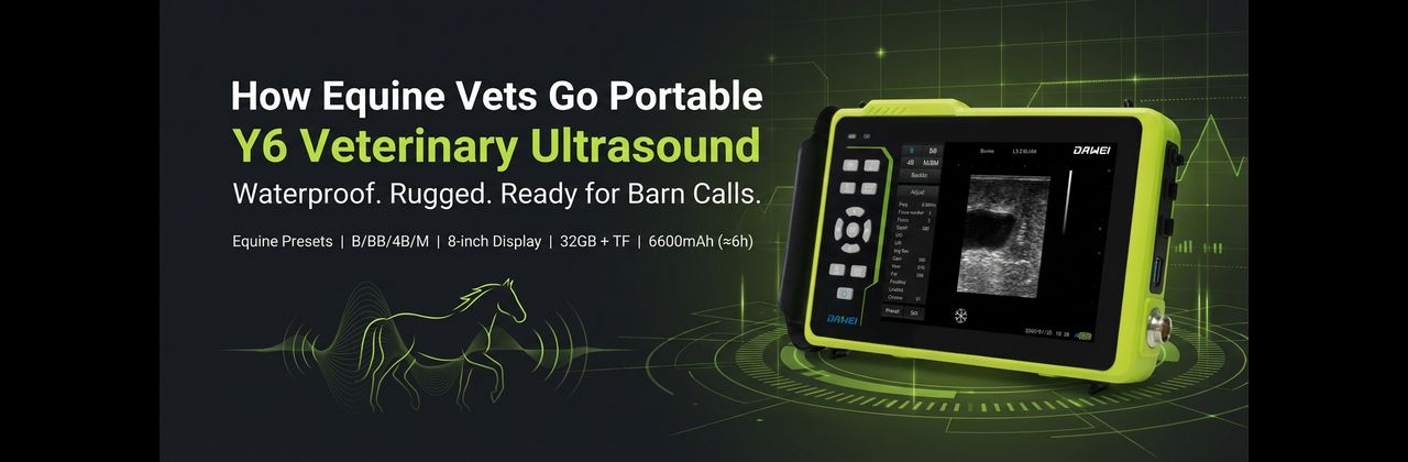
Where Y6 fits naturally in equine field work
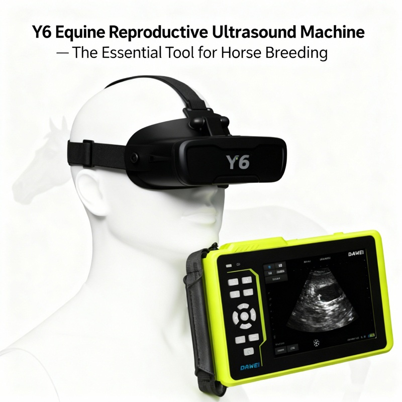
If you routinely scan horses outdoors or in wet/dirty conditions, a rugged, portable system can be a major advantage. For example, the Y6 Full Waterproof Veterinary Ultrasound Diagnostic System is designed for multi-species use (including equine) and supports common scan modes like B, BB, 4B, and M/BM.
From the provided specification, Y6 also includes:
- An 8-inch display and Chinese/English interface
- Presets for canine, feline, ovine, swine, bovine, equine
- 32GB storage with TF card expansion support
- Cine playback (255 frames)
- A 6600mAh battery rated for around 6 hours continuous operation (spec)
For equine practices, these points map well to real barn-call needs: quick documentation, portable power, and a workflow that supports frequent follow-up comparisons.
7) Durability: waterproofing, cleaning, and biosecurity
Equine ultrasound systems face:
- Dust, rain, mud, sweat
- Disinfectants and frequent wiping
- Travel vibration and accidental knocks
If you scan in unpredictable environments, prioritize durable housing, sealed buttons, and designs that are easy to clean between horses.
8) Matching the machine to common equine exams
Here are simple “best fit” matches you can use while comparing quotes:
- Equine tendon ultrasound: high-frequency linear probe, strong near-field resolution, easy measurements
- Equine abdominal ultrasound: low-frequency convex probe, good penetration, wide view
- Equine reproductive ultrasound:rectal probe option, fast preset switching, consistent grayscale
- Linear probe for MSK (tendons/ligaments)
- Convex probe for abdominal workups
- Rectal probe for repro (optional depending on caseload)
A practical probe configuration (example)
The Y6 spec supports probe types including convex, linear, rectal convex, and rectal linear scanning, which can simplify standardizing your equine toolkit across cases.
9) Total cost of ownership: probes, service, and training
When comparing “portable equine ultrasound price” quotes, include:
- Probe count and probe warranty terms
- Turnaround time for repairs
- Consumables and accessories (gel, sleeves, straps, cases)
- Training resources and presets
A slightly higher upfront cost can be worth it if downtime is lower during breeding season or a busy sport-horse calendar.
Frequently asked questions (FAQ)
What is the best ultrasound probe for horse tendon imaging?
Most veterinarians prefer a high-frequency linear probe for equine tendon ultrasound because tendons and ligaments are superficial and benefit from higher resolution.
Do I need a rectal probe for equine reproduction?
If your practice performs breeding soundness exams, follicle monitoring, or pregnancy checks, a transrectal ultrasound setup (rectal probe) is strongly recommended.
Is a waterproof veterinary ultrasound worth it for equine work?
For field-heavy equine practices, waterproofing and ruggedness can reduce failures caused by rain, wash-down areas, and repeated disinfection. It’s often a practical upgrade.
Conclusion: choose for your cases, then your environment
To choose the right portable veterinary ultrasound for equine imaging, start with your most common scans (tendon, abdomen, repro), pick the probes that match those exams, and then prioritize field-friendly features like battery life, durability, cine loops, and documentation.
If you want a field-ready option that supports equine presets and common modes (B/BB/4B/M), the Y6 full waterproof veterinary ultrasound system is a strong candidate to evaluate—especially for equine teams that split time between clinic and barn calls.
Post time: Feb-25-2026