


A customer case showing how the L3 laptop animal ultrasound machine system supports animal imaging, reproductive observation, and image data management at Changchun Institute of Applied Chemistry, Chinese Academy of Sciences.
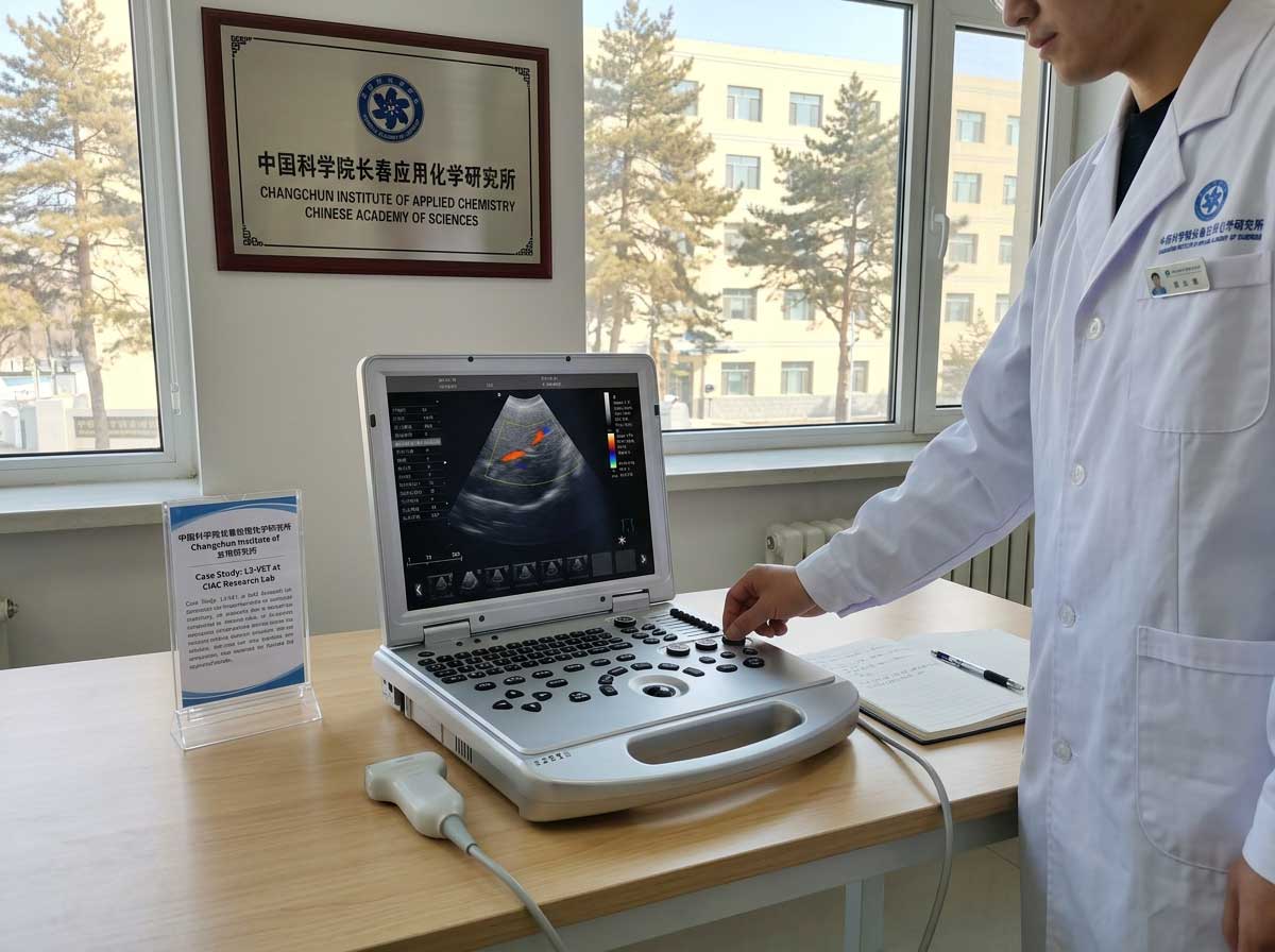
Customer Case: Changchun Institute of Applied Chemistry, Chinese Academy of Sciences
Product Used
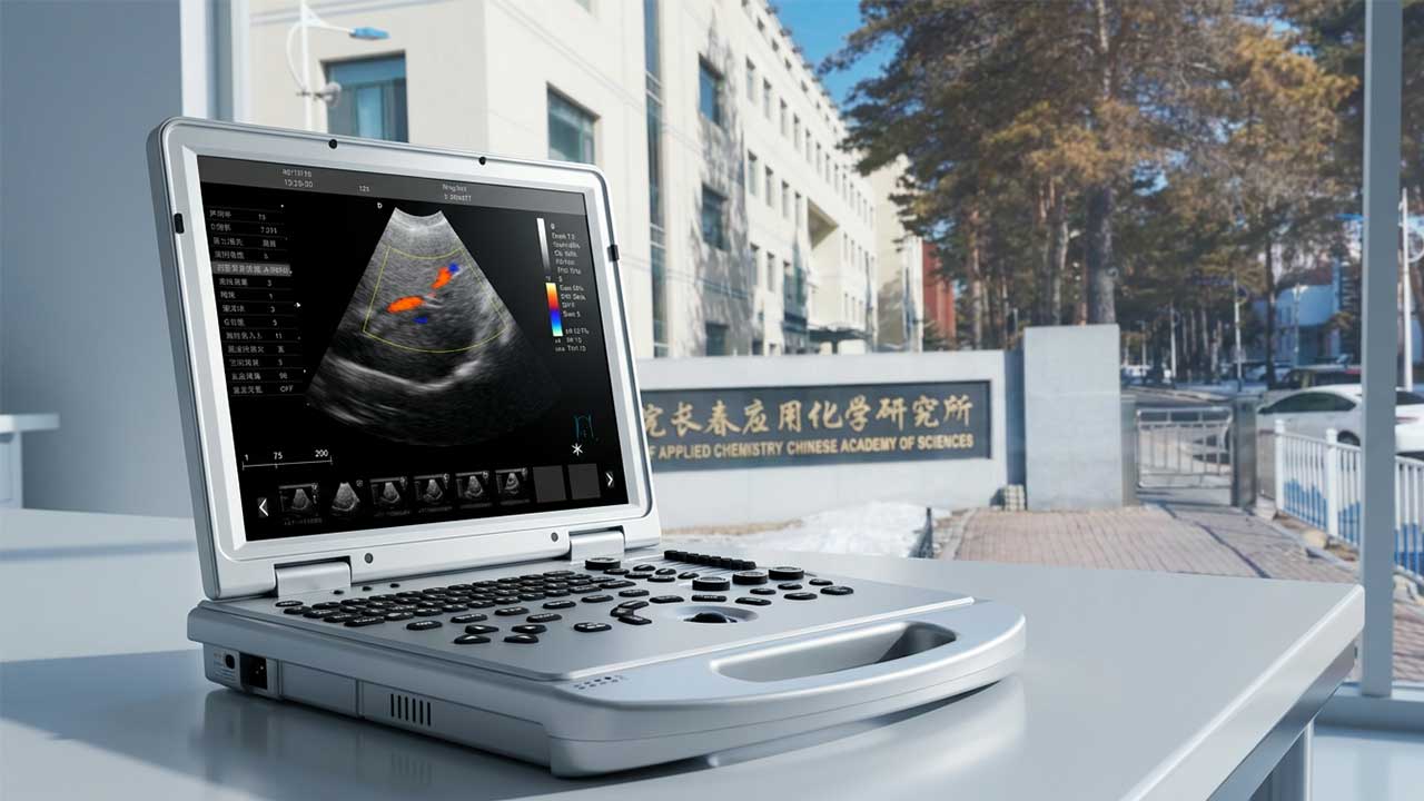
L3 Veterinary Color Doppler Ultrasound System
Project Location
Changchun, Jilin, China
Application Scenario
Scientific research unit / laboratory animal imaging / veterinary ultrasound examination
Project Background
As a research-oriented institution, the Changchun Institute of Applied Chemistry, Chinese Academy of Sciences requires imaging equipment that can fit into a structured laboratory workflow rather than simply perform as a standalone device. In practical use, the system needed to support routine ultrasound observation of research animals, assist with breeding- and reproduction-related examination, and provide clear image documentation for internal review and follow-up analysis.
Before adopting the L3, the institute was looking for a solution that could be used more flexibly across different working areas without creating a heavy operational burden for the research team. In a scientific research setting, imaging tasks are often spread across routine screening, scheduled observation, project-based examinations, and repeated follow-up checks. This means the ultrasound system must be quick to deploy, easy to move when needed, and stable enough for repeated daily use.
Another important consideration was workflow continuity. Research teams do not only need to obtain images during the examination itself; they also need to review, compare, store, and manage those images afterward. For that reason, the customer required a system that could combine practical imaging performance with user-friendly operation and manageable data handling.
Against this background, the L3 Veterinary Color Doppler Ultrasound System presented a strong match. Its laptop-style structure, Doppler capability, probe flexibility, and image management functions made it suitable for a research environment where portability, image quality, and documentation all matter.
Project Challenges
Although the institute’s application scenario was highly professional, the day-to-day challenges were very practical.
1. Different Examination Tasks Required More Than Basic Imaging
The customer’s use scenario was not limited to one single type of scan. Depending on the project stage and animal condition, the team could require routine abdominal observation, soft-tissue assessment, reproductive examination, or blood-flow related reference imaging. A basic black-and-white ultrasound system would not be flexible enough for these changing needs, so the customer needed a platform with broader imaging capability.
2. Equipment Had to Fit Into Real Laboratory Workflow
In research environments, it is rarely convenient to rely on bulky equipment that is difficult to move or slow to start up. The team needed a system that could be taken into different rooms or examination areas when necessary, set up quickly, and used without excessive adjustment time. If the operation process is too complicated, it can interrupt the rhythm of daily research work.
3. Different Animal and Examination Scenarios Called for Probe Flexibility
A single probe cannot efficiently cover all use scenarios in veterinary and research imaging. The customer needed the ability to adapt the system to different scanning tasks, whether for general examination, more focused observation, or reproduction-related applications. Probe compatibility was therefore an important part of the selection logic.
4. Image Records Needed to Be Easier to Store and Review
For a scientific research institute, examination data does not end with the scan itself. Images often need to be revisited for case review, internal discussion, comparative observation, or project documentation. A system without practical storage and data management functions would create extra manual work after the examination. The customer needed a solution that could support more organized image archiving and retrieval.
Why the L3 Was a Good Fit
The L3 is a laptop-style full-digital veterinary color Doppler ultrasound system designed for pet hospitals, clinics, zoos, breeding and reproduction bases, and scientific research units. For a professional research environment like Changchun Institute of Applied Chemistry, its value lies in combining portability, imaging capability, and data management in one system.
Key product features that make the L3 suitable for this kind of customer include:
- Portable laptop-type design that is easier to deploy across different rooms or work areas
- 15-inch display for clearer on-screen observation during examination
- Color Doppler and Pulsed Wave Doppler capability for blood-flow related assessment
- Tissue Harmonic Imaging (THI) and Compound Imaging for improved image optimization
- B/C/D real-time three synchronous imaging for more comprehensive observation
- Support for multiple probe options, including convex, linear, micro-convex, and rectal probes
- Built-in 128GB hard disk for fast and stable file storage
- DICOM 3.0 compatibility for integration with PACS-style image management workflows
- Portable form factor with built-in battery support for flexible use where mobility is important
According to the supplementary product information, the L3 also offers a compact structure, with a size of approximately 375 × 360 × 75 mm, a weight of about 6.7 kg, and a battery capacity of 15600 mAh, supporting approximately 3 to 6 hours of use depending on the operating condition.
What the L3 Can Help the Institute Do
In a scientific research organization such as Changchun Institute of Applied Chemistry, the L3 can support a range of practical imaging tasks.
1. Support Routine Animal Examination and Screening
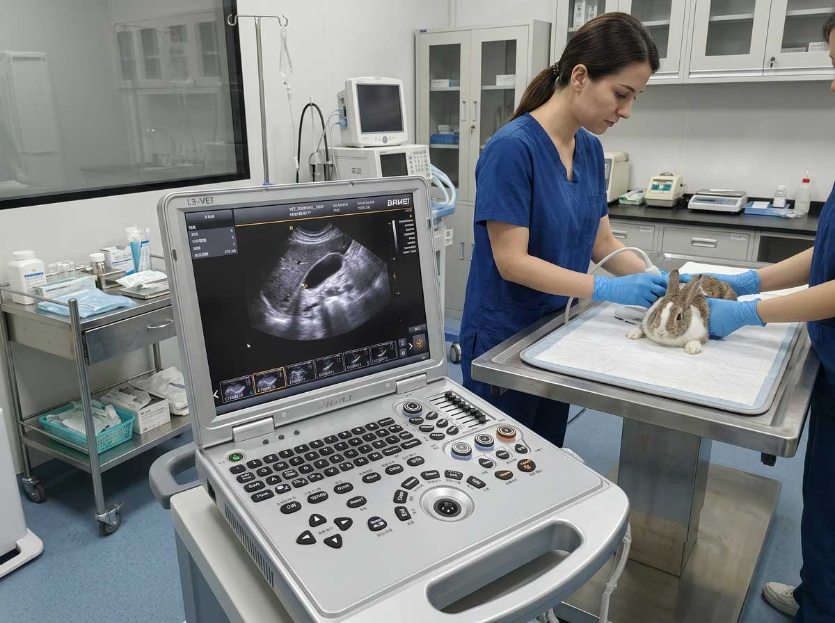
The L3 can be used for routine ultrasound examination of research animals and veterinary subjects, helping teams carry out non-invasive observation more efficiently. Its portable structure makes it suitable for use in laboratories, animal rooms, and other controlled research spaces where flexibility and convenience are important.
2. Assist Reproductive and Breeding-Related Observation
With optional micro-convex and rectal probes, the system can support reproductive and breeding-related ultrasound applications. For research teams involved in animal breeding, reproductive assessment, or developmental observation, the L3 provides a more practical imaging tool for regular checks and follow-up.
3. Enable Soft Tissue, Abdominal, and Organ Assessment
The system includes professional measurement and analysis packages for different organs, including abdomen and OB-related applications. This makes it useful for abdominal observation, soft tissue evaluation, and general organ-oriented imaging tasks in veterinary and research settings.
4. Provide Blood Flow Information When Needed
Because the L3 supports color Doppler, directional power Doppler imaging, and pulsed wave Doppler, it can assist users in observing blood-flow related information in relevant examination scenarios. This expands the system’s usefulness beyond basic black-and-white imaging and gives research users more diagnostic reference dimensions.
5. Improve Research Data Documentation and Review
For institutes that care about documentation and traceability, the L3 offers practical file and record management functions. The system can store image and patient information locally, supports multiple image export formats such as BMP, DCM, and JPG, and can connect to PACS through DICOM 3.0. This helps improve internal archiving, retrieval, and review efficiency.
Implementation Process
In a use environment like this, the value of the L3 is reflected not only in specifications, but also in how smoothly it can be integrated into actual work.
First, the system can be deployed as a mobile imaging unit for routine examination and project-based observation. Because of its laptop-style structure and battery-supported operation, the device is easier to bring into the required working area instead of forcing every examination to happen around a fixed station.
Second, the research team can match the probe configuration to the task at hand. General examination can be handled with standard probes, while more specific applications such as reproductive observation or focused anatomical assessment can be supported through appropriate optional probes. This makes the system more adaptable to real-world scientific use rather than limiting it to a narrow examination range.
Third, built-in optimization functions and preset capabilities help reduce repeated manual adjustment during routine use. For teams that need to perform recurring examinations, this can improve consistency and make the workflow easier for operators to standardize.
Finally, once the examination is complete, the system supports image review, storage, export, and downstream record management. This is especially valuable in research settings where image documentation may need to be checked again later rather than used only once at the point of care.
Project Results
After integrating the L3 into the imaging workflow, the customer gained a more practical and efficient examination setup for research-related use.
More Flexible Day-to-Day Operation
The portable form factor made it easier to use the system in the actual working environment instead of treating ultrasound examination as a fixed-location task only. This improved operational flexibility and reduced the inconvenience commonly associated with larger, less mobile systems.
Better Coverage Across Multiple Use Scenarios
With support for multiple imaging modes and optional probes, the L3 was better aligned with the institute’s varied examination needs. Instead of depending on one limited setup, the team could use the same platform across routine checks, reproductive observation, abdominal examination, and selected blood-flow related applications.
More Efficient Examination Workflow
The combination of one-key smart optimization, presets, clear display, and integrated workflow functions helped make scanning more straightforward in daily use. For the customer, this meant less time spent on repeated adjustment and a smoother path from examination to image review.
Stronger Image Documentation and Traceability
The built-in storage, export formats, and DICOM/PACS compatibility improved the practicality of post-examination data handling. For a research institution, this is a meaningful result because it supports more organized image retention, internal sharing, and follow-up comparison.
Expected Benefits for the Customer
In a research-driven use environment, the L3 can help deliver several practical benefits:
- More convenient deployment for daily imaging tasks
- Better adaptability across multiple examination scenarios
- Improved imaging support for reproductive, abdominal, and blood-flow observation
- Smoother image storage, review, and record management
- Greater workflow efficiency through portable design and smart operation features
Customer-Oriented Summary
For institutions such as Changchun Institute of Applied Chemistry, Chinese Academy of Sciences, ultrasound equipment is not simply a device purchase. It is part of a broader research and examination workflow. The L3 addresses this need by combining mobility, imaging capability, probe flexibility, and data management in one system.
Whether used for routine animal screening, breeding-related observation, organ examination, or research image documentation, the L3 provides a practical and professional solution for scientific research units that require both performance and usability.
Technical Highlights of the L3
- Laptop-type veterinary color Doppler ultrasound system
- 15-inch monitor
- PW Doppler, DPDI, THI, and Compound Imaging
- B/C/D real-time three synchronous imaging
- 2B/4B imaging modes
- One-key smart optimization
- Optional convex, linear, micro-convex, and rectal probes
- Built-in 128GB hard disk
- Image export in BMP, DCM, and JPG formats
- DICOM 3.0 / PACS connectivity
- Approx. 6.7 kg weight and 3–6 hours battery support
Conclusion
This case reflects how the L3 Veterinary Color Doppler Ultrasound System can serve the needs of a professional scientific research institution. With its portable design, advanced imaging functions, and practical data management features, the L3 can support more efficient and flexible ultrasound workflows in research and veterinary application scenarios.
For customers looking for a compact yet capable veterinary ultrasound solution for research units, breeding bases, clinics, or specialized animal examination environments, the L3 offers a strong balance of usability, functionality, and workflow value.
Post time: Jun-26-2026