


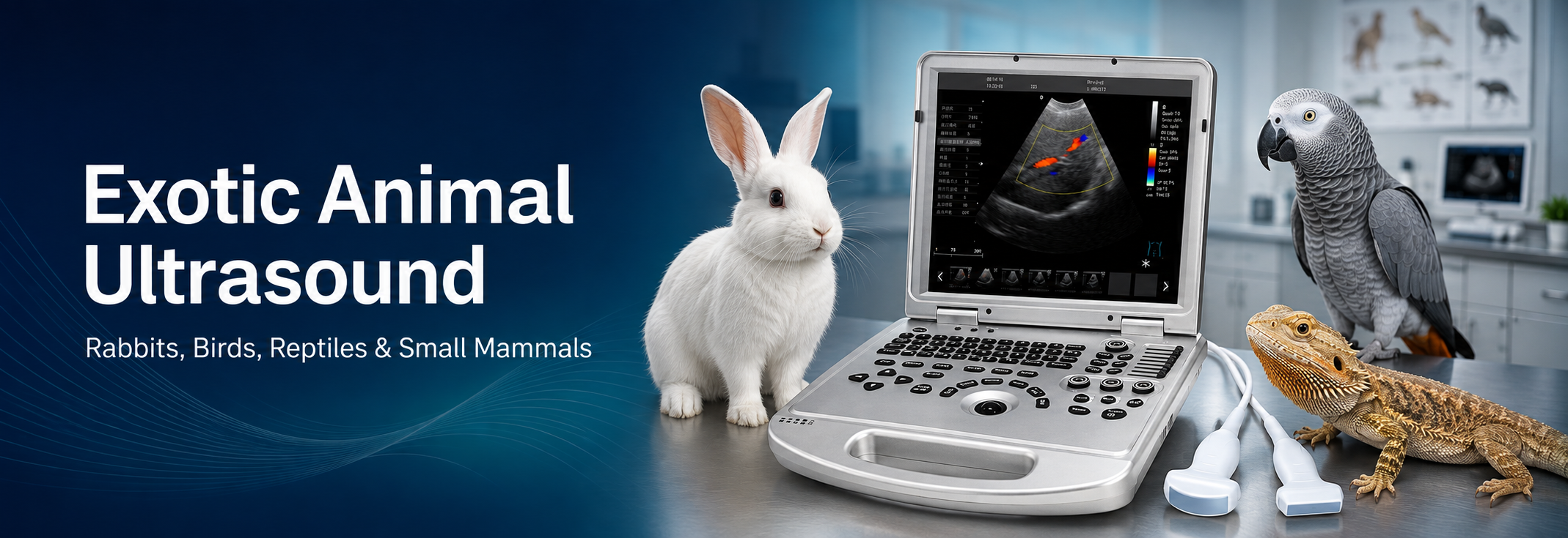
Exotic Animal Ultrasound: Imaging Rabbits, Birds, Reptiles, and Small Mammals
Ultrasound is no longer reserved for dogs, cats, and large animals. In an exotic-animal practice, it can provide real-time information about the abdomen, heart, reproductive tract, urinary system, superficial tissues, and selected masses without exposing the patient to ionizing radiation. That matters when the patient is a 1.5-kilogram rabbit, a fragile parrot, or a lizard whose anatomy does not resemble that of a mammal.
The appeal is obvious. The execution is not.
Exotic animal ultrasound demands a small acoustic footprint, carefully selected frequency, gentle restraint, and a working knowledge of species-specific anatomy. A preset that produces a useful canine abdominal image may be poorly suited to a cockatiel or bearded dragon. Even within one group, body size, feathers, scales, shell, gastrointestinal gas, air sacs, temperature, and stress response can change the examination.
This guide explains how veterinary teams can approach ultrasound imaging in rabbits, birds, reptiles, ferrets, guinea pigs, rodents, and other small mammals. It also shows how probe choice and system capabilities affect what the operator can realistically see.
Quick Answer: Can Ultrasound Be Used on Exotic Animals?
Yes. Veterinary ultrasound can be used on many exotic species for abdominal, reproductive, cardiac, urinary, vascular, ocular, and superficial soft-tissue assessment. The best results usually come from a high-frequency linear or small-footprint microconvex probe, although the ideal probe depends on patient size, target depth, and available acoustic window.
Ultrasound should not be treated as a universal replacement for radiography, CT, MRI, endoscopy, or laboratory testing. Gas, air sacs, bone, shells, and very small structures can limit the examination. In practice, ultrasound is strongest when the clinical question is specific: Is free fluid present? Is the urinary bladder distended? Does a hepatic lesion have internal blood flow? Is the reproductive tract enlarged? Is a mass solid, cystic, or mixed?
Why Exotic Animal Ultrasound Is Different
An exotic patient is not simply a smaller dog. Several variables change both image acquisition and interpretation.
Small anatomy requires higher spatial resolution
When the organ itself measures only a few millimeters, small changes in focal position, gain, depth, and probe pressure become important. Higher ultrasound frequencies generally improve detail but reduce penetration. The operator therefore needs enough frequency for the target structure without sacrificing the depth required to reach it.
The acoustic window may be narrow
Feathers trap air. Reptile scales and keratinized structures can weaken contact. Air sacs in birds and gastrointestinal gas in rabbits may block deeper organs. Chelonians add a shell. The examination often depends on finding a small, repeatable window rather than sweeping across a broad clipped abdomen as one might in a dog.
Restraint is part of image quality
Excessive pressure can distort small organs, alter blood flow, or compromise breathing. Prolonged handling also increases stress and motion. Some patients tolerate conscious scanning with skilled restraint; others may require sedation or anesthesia based on species, condition, and the diagnostic objective. That decision belongs to the attending veterinarian and must account for anesthetic risk.
Normal anatomy varies sharply by species
A rabbit has a large gastrointestinal tract that can dominate the image. A bird has air sacs and no diaphragm separating thorax and abdomen in the mammalian sense. Reptilian cardiac anatomy, position, and blood-flow patterns differ substantially from those of mammals. Reference images and measurements should therefore be species-specific whenever possible.
Rabbit Ultrasound: A Practical Starting Point
Rabbit ultrasound is one of the most useful entry points into exotic imaging because the abdomen can often be examined transcutaneously with a compact probe. Common clinical questions involve the liver, kidneys, urinary tract, gastrointestinal tract, reproductive system, free abdominal fluid, and abdominal masses.
A focused abdominal study published in 2024 used a 3-11 MHz microconvex probe in conscious rabbits positioned in sternal recumbency. Through xiphisternal, bilateral renal, and cystic windows, the investigators consistently identified the liver, kidneys, stomach, duodenum, jejunum, caecum, and colon. The bladder, gallbladder, spleen, and major vessels were also visible in many animals. The important lesson is not that one protocol fits every rabbit. It is that a small-footprint probe and a limited set of deliberate windows can produce a surprisingly useful examination.
Common rabbit ultrasound applications
- · Hepatobiliary assessment, including liver echogenicity, focal lesions, gallbladder appearance, and perfusion when Doppler is indicated.
- · Renal and urinary evaluation for kidney architecture, pelvic dilation, bladder contents, sediment, uroliths, obstruction, or masses.
- · Reproductive imaging of the uterus, ovaries, pregnancy, retained fluid, wall changes, or suspected uterine disease.
- · Gastrointestinal evaluation for distension, wall appearance, motility, focal masses, fluid, and the relationship between affected bowel and surrounding tissues.
- · Detection of abdominal effusion, abscesses, enlarged lymph nodes, and other space-occupying lesions.
- · Focused cardiac or vascular examination when a suitable probe and species-appropriate protocol are available.
Rabbit gastrointestinal gas and ingesta can hide deeper anatomy, so a negative or incomplete scan should not be overinterpreted. Changing the patient’s position, reducing depth, using a different window, or repeating the scan after stabilization may help. Ultrasound findings should be interpreted alongside history, physical examination, radiographs, laboratory results, and, when necessary, advanced imaging.
For laboratories working with much smaller rodents, the equipment and protocol may be different. Our overview of veterinary ultrasound systems for laboratory mouse examination discusses reproductive monitoring, organ assessment, disease models, and longitudinal research applications.
Avian Ultrasound: Working Around Feathers and Air Sacs
Avian ultrasound is valuable precisely because birds present diagnostic challenges. Radiography remains central to avian imaging, but ultrasound can clarify whether a coelomic opacity is solid or fluid-filled, characterize effusion, examine accessible portions of the liver and heart, evaluate reproductive abnormalities, and guide selected procedures.
The main obstacle is air. Feathers and air sacs interfere with sound transmission, while the small body size of many birds leaves little room for probe manipulation. The operator may use a ventromedian, parasternal, lateral, or intercostal window depending on the species and target. Feathers are parted, and a generous amount of warmed acoustic gel can improve coupling. Clipping is sometimes unnecessary and may be undesirable, but contact must be reliable.
What avian ultrasound may help assess
- · Coelomic effusion and the relationship of fluid to adjacent organs.
- · Liver size, margins, echogenicity, focal changes, and selected vascular features.
- · Cardiac motion and gross chamber or flow assessment when a suitable window is available.
- · Reproductive tract abnormalities, including enlarged follicles, eggs, masses, or fluid-filled structures.
- · Ocular disease and selected superficial lesions with a high-frequency probe.
- · Image-guided aspiration or biopsy in carefully selected cases.
Birds have limited respiratory reserve, so patient positioning and probe pressure deserve particular care. The sternum must be allowed to move. If restraint impairs ventilation, the examination has already become unsafe, regardless of image quality.
Published avian work demonstrates that ultrasound is more than a screening tool. Studies have used it to guide liver biopsy, examine the eye, and characterize coelomic disease. Still, image interpretation can be difficult because normal organ visibility changes with species, body condition, air-sac inflation, reproductive status, and disease. A clean image is not automatically a complete examination.
Reptile Ultrasound: Anatomy, Temperature, and Species Matter
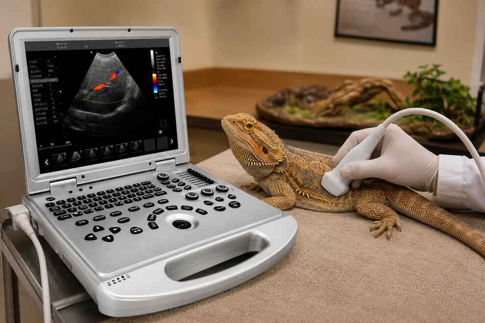
Reptile ultrasound has expanded from reproductive checks to coelomic, cardiac, vascular, hepatic, renal, and soft-tissue imaging. It can be especially helpful in lizards and snakes, where the probe may access the coelom through a relatively broad soft-tissue surface. Turtles and tortoises are more difficult because the carapace and plastron restrict the acoustic window; scanning may depend on prefemoral or cervical soft-tissue windows.
Before scanning, the operator should understand the animal’s normal body temperature range, recent feeding status, and anatomy. Reptilian physiology is temperature dependent, and heart rate or gastrointestinal activity may be misleading if the patient is outside an appropriate thermal range. The position of the heart, kidneys, gonads, and fat bodies also varies among snakes, lizards, and chelonians.
Common reptile ultrasound applications
- · Reproductive assessment, including follicles, eggs, testes, and suspected egg retention.
- · Liver, gallbladder, kidney, urinary bladder, and fat-body evaluation where anatomy and windows permit.
- · Detection of coelomic fluid, abscesses, granulomas, cysts, and solid masses.
- · Cardiac imaging and Doppler assessment in species for which the operator has appropriate reference knowledge.
- · Guidance for fluid sampling, biopsy, or other interventions when clinically justified.
Snakes illustrate why mammalian assumptions can fail. The ophidian heart is mobile within the coelom, and its ventricular anatomy and outflow tracts are distinct. A study of healthy ball pythons showed that two-dimensional and pulsed-wave Doppler imaging could document cardiac structures and blood flow, but interpretation required an understanding of reptilian circulation. Simply applying canine cardiac measurements would be inappropriate.
For routine clinical work, a microconvex probe often provides a useful balance of footprint and field of view. A high-frequency linear probe can add detail for superficial organs, small patients, vessels, and musculoskeletal lesions. Color and spectral Doppler are useful when the question involves perfusion, vascular patency, or cardiac flow, but low-flow settings and careful pressure control are essential.
Ultrasound in Ferrets, Guinea Pigs, Hamsters, and Other Small Mammals
The phrase “small mammals” covers animals with very different anatomy. Ferrets may be scanned with techniques familiar from cats, although their elongated body shape and species-specific diseases change the differential list. Guinea pigs and chinchillas have large, gas-filled gastrointestinal tracts that can obstruct the acoustic path. Hamsters, mice, gerbils, hedgehogs, sugar gliders, and other small patients push the limits of probe footprint, frequency, and operator hand control.
Common applications include:
- · Abdominal organ assessment, particularly the liver, kidneys, spleen, bladder, and reproductive tract.
- · Investigation of abdominal distension, reduced appetite, weight loss, urinary signs, or a palpable mass.
- · Pregnancy and reproductive tract evaluation where ultrasound is clinically indicated.
- · Cardiac screening or targeted echocardiography in patients large enough for the available probe and validated protocol.
- · Superficial soft-tissue, ocular, vascular, or musculoskeletal imaging.
- · Follow-up of a known lesion without repeated exposure to ionizing radiation.
Very small rodents used in research may need ultra-high-frequency systems for fine cardiac or developmental measurements. A conventional veterinary color Doppler system can still support broader abdominal, reproductive, or vascular questions in selected small mammals, but it should not be marketed as equivalent to a dedicated preclinical micro-ultrasound platform. The clinical question must match the system’s true resolution.
Which Probe Is Best for Exotic Animal Ultrasound?
There is no single “exotic probe.” The best choice is the one that places the target within the focal zone, provides enough penetration, fits the available acoustic window, and can be held without excessive pressure.
| Probe type | Main advantage | Typical exotic-animal uses | Important limitation |
| Microconvex | Small footprint with a broad sector image | Rabbit abdomen, small mammal abdomen, reptile coelom, focused scans | Less superficial detail than a high-frequency linear probe |
| High-frequency linear | Excellent near-field detail and rectangular image | Superficial organs, vessels, eye, soft tissue, small patients, image-guided procedures | Wider footprint and limited penetration can restrict access |
| Phased array | Very small contact area and sector image | Selected cardiac examinations through narrow windows | Cardiac interpretation is highly species dependent |
| Convex | Wider field and greater penetration | Larger rabbits, large reptiles, deeper coelomic or abdominal targets | Footprint may be too large for small birds and small mammals |
Frequency should be adjusted rather than treated as a fixed number. Start high enough to resolve the structure, then reduce frequency only if penetration is inadequate. Depth should be shallow enough that the organ occupies a useful portion of the screen. Set the focal zone at or just below the structure of interest. Use gain to reveal tissue texture without turning fluid gray or erasing subtle boundaries.
A Safer, More Repeatable Scanning Workflow
1. Define the clinical question
“Scan the abdomen” is broad. “Look for free fluid and assess the liver and urinary bladder” is actionable. A clear question determines the patient position, probe, preset, and time required.
2. Stabilize the patient first
Correct hypothermia, severe respiratory distress, shock, or pain as appropriate before extending the examination. Imaging should not delay urgent care.
3. Select the smallest suitable probe
Use a microconvex probe when access is tight but deeper structures must be seen. Use a high-frequency linear probe for superficial detail. For cardiac work, select a probe that fits the window and is supported by a species-appropriate protocol.
4. Prepare the contact surface gently
Part fur or feathers, remove trapped air, and use warmed gel. Clipping may improve contact in some mammals, but the decision should consider owner preference, feather integrity, species, and urgency. Do not use excessive alcohol or cold gel on fragile or hypothermic patients.
5. Minimize pressure and examination time
Rest the hand against the table or patient support to make small movements. Reduce probe pressure when using color Doppler or examining delicate patients. Save representative still images and cine loops as the examination proceeds so that useful information is not lost if the patient becomes stressed.
6. Document limitations
State which organs were seen, which were only partially evaluated, and what prevented complete assessment. Gas, shell, air sacs, patient motion, and lack of an appropriate probe are meaningful limitations, not footnotes.
Common Mistakes in Exotic Animal Ultrasound
Using a probe that is physically too large
A high-resolution system cannot compensate for a footprint that does not fit the acoustic window. If only part of the probe contacts the patient, the image will be unstable and misleading.
Scanning too deep
Exotic patients often occupy only the top few centimeters of the image. Excessive depth makes small structures tiny and wastes lines of sight. Reduce depth early.
Applying too much pressure
Pressure can compress veins, displace fluid, deform soft organs, and restrict respiration. This is particularly important in birds and very small mammals.
Assuming one species’ normal is another species’ normal
Measurements and appearance must be interpreted using appropriate references. A normal ball python heart does not follow canine conventions; a bird’s coelomic anatomy does not map neatly onto a cat.
Overpromising what ultrasound can diagnose
Ultrasound findings often narrow the problem rather than produce a final diagnosis. Histopathology, cytology, CT, radiography, endoscopy, or laboratory testing may still be required. Product marketing should reflect this clinical reality.
How to Choose an Ultrasound Machine for Exotic Animals
An effective ultrasound machine for exotic animals must do more than produce a bright image. It should support the probe range, settings, and workflow needed for small, easily stressed patients.
Consider the following:
- · Probe compatibility: Confirm availability of microconvex, high-frequency linear, convex, and phased-array probes according to your caseload.
- · Near-field image quality: Small organs frequently lie close to the skin, so superficial resolution and focal control matter.
- · Fast optimization: One-key optimization, responsive controls, and useful presets can shorten handling time.
- · Doppler sensitivity: Color and spectral Doppler may be important for vascular, perfusion, reproductive, and cardiac questions.
- · Cine-loop and storage: Exotic patients move. Frame-by-frame review, still-image capture, video storage, and export help preserve the diagnostic moment.
- · Portability: A laptop-style or compact system can move between exam rooms, wards, research areas, and mobile settings.
- · Display and ergonomics: The operator must see subtle structures while maintaining gentle restraint and precise probe control.
- · Training and service: Probe selection, presets, image optimization, and species-specific training influence the value of the equipment after purchase.
Clinics comparing systems can browse Dawei’s Pet Ultrasound Machines category. For practices that need abdominal, Doppler, and mobile imaging support across companion animals and selected exotics, the P5S-VET portable veterinary Doppler ultrasound is designed for veterinary hospitals, specialty clinics, mobile services, and animal research settings. Final probe configuration should always be confirmed against the species, examination target, and local quotation.
For a different research scenario, see how the P3-VET supports non-invasive observation and documentation in an aquatic research and breeding customer case.
Frequently Asked Questions About Exotic Animal Ultrasound
What is exotic animal ultrasound used for?
Exotic animal ultrasound is used to evaluate accessible internal organs, reproductive structures, the urinary system, the heart, blood flow, superficial tissues, masses, and free fluid in species such as rabbits, birds, reptiles, ferrets, guinea pigs, rodents, and zoo or wildlife patients. The exact application depends on anatomy, body size, acoustic window, probe selection, and operator experience.
What is the best probe for rabbit ultrasound?
A small-footprint microconvex probe is often a practical first choice for rabbit abdominal imaging because it provides a sector field through a narrow contact area. A high-frequency linear probe can improve detail in superficial structures. The final frequency and probe depend on rabbit size, organ depth, and the clinical question.
Can ultrasound be used on birds?
Yes. Avian ultrasound can assist with coelomic fluid, liver changes, selected cardiac and reproductive findings, ocular disease, masses, and image-guided procedures. Feathers and air sacs limit transmission, so the operator must find an appropriate acoustic window and avoid restricting sternal movement.
Can a veterinarian perform ultrasound on reptiles?
Yes. Ultrasound is used in reptiles for reproductive, coelomic, cardiac, hepatic, renal, vascular, and soft-tissue assessment. Access is generally easier in snakes and many lizards than in chelonians, where the shell limits the window. Interpretation must account for species-specific anatomy and temperature-dependent physiology.
Is sedation always required for exotic animal ultrasound?
No. Some rabbits, reptiles, birds, and small mammals can be scanned while conscious with skilled, gentle restraint. Sedation or anesthesia may be necessary when stress, motion, pain, procedure type, or patient safety prevents a useful examination. The decision should be made by the attending veterinarian for the individual patient.
Is ultrasound safe for very small animals?
Diagnostic ultrasound does not use ionizing radiation and is widely used for repeat imaging. Safe practice still requires appropriate output settings, short examination times, gentle pressure, thermal awareness, and monitoring of fragile patients. The lowest settings that answer the clinical question are a sensible principle.
Can one veterinary ultrasound machine scan every exotic species?
One versatile system may cover many clinical applications when it supports multiple probes and flexible settings. It may not replace specialized high-frequency research ultrasound for extremely small structures or every advanced imaging modality. Buyers should match the system, probes, presets, and training to their actual species and diagnostic goals.
What should a clinic ask before requesting a quotation?
Prepare a list of the species most often examined, the smallest and largest patient sizes, the organs or applications required, whether cardiac or Doppler imaging is needed, and whether the system will be used in a fixed clinic or mobile setting. This information helps the supplier recommend a more appropriate machine and probe package.
Final Takeaway
Exotic animal ultrasound succeeds when the examination is adapted to the patient. Rabbits often benefit from a compact abdominal protocol. Birds require careful respiratory handling and small acoustic windows. Reptiles demand species-specific anatomical knowledge, while very small mammals push the limits of frequency, footprint, and hand control.
The machine matters. The probe matters more than many buyers expect. Yet the largest difference comes from matching the clinical question, patient preparation, acoustic window, system settings, and operator training.
If your clinic, zoo, research institute, or mobile veterinary service is evaluating ultrasound for rabbits, birds, reptiles, or small mammals, contact Dawei with your target species and examination needs. Our team can help you compare suitable veterinary ultrasound systems and probe configurations before you request a quotation.
Post time: Aug-11-2026