


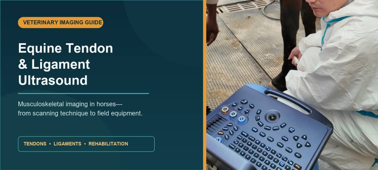
Quick answer: Equine tendon and ligament ultrasound is a non-invasive diagnostic imaging examination used to assess soft tissues in a horse’s limb. In real time, it can help a veterinarian evaluate tendon or ligament size, shape, fiber pattern, echogenicity, margins, and surrounding fluid. It is particularly useful for suspected flexor tendon and suspensory ligament injuries, as well as for documenting change during a veterinarian-directed rehabilitation program.
One caveat matters from the outset: diagnostic ultrasound does not produce a verdict by itself. The scan must be interpreted alongside the horse’s history, palpation findings, lameness examination, diagnostic analgesia when appropriate, and—sometimes—radiographs, MRI, CT, or other imaging.
What Does Equine Musculoskeletal Ultrasound Show?
Ultrasound sends high-frequency sound waves into tissue and translates the returning echoes into a live grayscale image. Bone surfaces reflect most of the beam. Soft tissues, however, can be examined in revealing detail—especially superficial, fiber-rich structures such as tendons and ligaments.
During an equine tendon ultrasound, the clinician commonly looks at:
- cross-sectional area and overall shape;
- echogenicity, or how bright and dark the tissue appears;
- the alignment and continuity of the fiber pattern;
- focal defects, irregular margins, or enlargement;
- fluid within or around a tendon sheath;
- change over time when serial scans are obtained with a consistent technique.
Normal tendon fibers tend to create a regular, parallel pattern in a longitudinal view and a more uniform texture in transverse section. A lesion may appear enlarged, darker, less organized, or irregular. Yet those words are not a diagnosis. Tendons and ligaments are strongly anisotropic: simply tilting the transducer away from a perpendicular angle can make healthy fibers look artificially dark.
That artifact is one reason technique matters so much. The American Association of Equine Practitioners describes how on-angle and off-angle imaging can help distinguish true abnormalities from angle-dependent changes, particularly within the suspensory ligament.
Which Tendons and Ligaments Can Be Examined?
The exact scan depends on where pain or swelling has been localized. In the distal limb, frequently evaluated structures include:
- the superficial digital flexor tendon (SDFT);
- the deep digital flexor tendon (DDFT);
- the accessory ligament of the DDFT, often called the inferior check ligament;
- the suspensory ligament, including its origin, body, and branches;
- distal sesamoidean ligaments;
- tendon sheaths and adjacent soft tissues.
Ultrasound is also used beyond the classic palmar or plantar metacarpal region. Depending on anatomy, access, and equipment, an equine veterinarian may assess soft tissues around the pastern, fetlock, carpus, hock, shoulder, stifle, pelvis, or back. Some regions are more challenging because they are deep, curved, hidden by bone, or difficult to approach safely.
The practical lesson is simple: “musculoskeletal ultrasound” covers a broad family of examinations. A scan optimized for a superficial flexor tendon is not automatically the right setup for a deeper structure.
When Might a Veterinarian Recommend Horse Ligament Ultrasound?
A veterinarian may recommend horse ligament ultrasound or tendon imaging when a horse has:
- localized heat, pain, swelling, or thickening;
- acute or persistent lameness localized to a soft-tissue region;
- an unexplained decline in performance;
- a suspected superficial digital flexor tendon injury;
- suspected suspensory ligament desmitis or branch injury;
- a tendon-sheath problem;
- a known lesion that needs scheduled reassessment.
Not every tendon injury presents with dramatic swelling. Subclinical microdamage can precede obvious clinical tendinopathy, particularly in athletic horses exposed to repetitive loading. B-mode ultrasonography remains the most commonly used modality for detecting and monitoring many metacarpal and metatarsal tendon lesions, according to a review of equine flexor tendon imaging.
Still, routine screening and clinical diagnosis are not the same thing. A dark area on a single image should never be divorced from probe angle, symmetry, anatomy, and the rest of the examination.
How Is an Equine Tendon and Ligament Ultrasound Performed?
Protocols vary by structure and by clinician, but a careful examination usually follows a repeatable sequence.
1. Localize the region of interest
The veterinarian first integrates history, observation at rest and in motion, palpation, flexion tests, hoof assessment, and diagnostic analgesia when indicated. Ultrasound works best when there is a well-defined anatomical question.
2. Prepare the scan site
Hair traps air, and air interferes with sound transmission. The area is therefore commonly clipped, cleaned, and wetted; ultrasound gel is applied to improve acoustic contact. In some circumstances a stand-off pad or a generous layer of gel helps with very superficial structures.
3. Position the horse and limb consistently
A quiet, level surface is important. The horse may be examined while weight-bearing, while the limb is briefly unloaded, or in both conditions, depending on the target. Safety for the horse, handler, and veterinarian governs every choice.
4. Scan in two orthogonal planes
The transducer is moved through sequential anatomical zones in transverse and longitudinal planes. Images in two planes help confirm whether an apparent abnormality is reproducible rather than an artifact. The opposite limb may be scanned for comparison—but it is a reference, not a guaranteed “normal control.” Bilateral disease exists.
5. Control beam angle and machine settings
Gain, depth, focus, frequency, and time-gain compensation should be adjusted so the target—not the surrounding empty space—receives the available resolution. For superficial flexor tendons and the suspensory ligament, the MSD Veterinary Manual notes that a 7.5–10 MHz linear probe is commonly suitable. Probe choice changes with depth and anatomy.
The clinician also “fans” or angles the beam deliberately. Because fiber echogenicity changes with angle, this small wrist movement can separate genuine fiber disruption from anisotropy.
6. Measure and archive
Relevant images, anatomical level, dimensions, and cross-sectional area can be recorded. Cine loops may preserve dynamic information. Good labeling and a consistent protocol become especially valuable during follow-up: the comparison is meaningful only when the same structure and level are being compared.
What Do Normal and Abnormal Findings Look Like?

| Feature | Common appearance in normal tissue | Finding that may warrant investigation |
|---|---|---|
| Size and shape | Expected dimensions and smooth contour for that anatomical level | Focal or diffuse enlargement, asymmetry, contour change |
| Echogenicity | Relatively uniform when the beam is correctly aligned | Persistent focal hypoechogenicity or mixed echogenicity |
| Longitudinal fiber pattern | Parallel, continuous linear echoes | Reduced alignment, interruption, or irregularity |
| Transverse pattern | Organized, repeatable texture | Focal defect or heterogeneous region seen in a second plane |
| Margins | Smooth and clearly defined | Irregular or poorly defined boundary |
| Adjacent tissues | No unexpected fluid or marked tissue reaction | Tendon-sheath fluid, edema, or surrounding change in context |
This table is descriptive, not diagnostic. Normal anatomy varies by zone, breed, age, discipline, limb loading, and individual. The proximal suspensory ligament is a classic trap because normal muscle and fat can produce a mottled appearance. A complete equine suspensory ligament ultrasound examination may therefore include on-incidence, off-incidence, weight-bearing, and non-weight-bearing views.
Color or power Doppler can add information about blood flow in selected cases. Elastography and ultrasound tissue characterization are also being studied as ways to quantify tissue properties. These techniques are promising, but conventional B-mode imaging remains the field workhorse, and advanced measurements should not be oversold. A 2025 review of quantitative imaging emphasizes that emerging ultrasound, CT, and MRI methods are complementary rather than interchangeable.
Can Ultrasound Grade the Severity of a Horse Tendon Injury?
Ultrasound can help describe lesion extent and location. Depending on the protocol, a veterinarian may document the percentage of a structure involved, cross-sectional area, fiber disruption, or lesion length. Those observations can contribute to prognosis and rehabilitation planning.
But severity is not a single grayscale number. A small lesion in a highly loaded location may matter greatly. A chronic tendon may look more echogenic while its mechanical properties remain altered. Scar tissue is not identical to the original hierarchical fiber architecture. And an apparently improved image does not, on its own, prove that the tissue is ready for full-speed work.
The scan answers structural questions. The veterinarian answers the clinical one: what does this mean for this horse, in this discipline, at this stage?
How Is Ultrasound Used to Monitor Healing?
Serial imaging can track change in lesion size, echogenicity, and fiber organization. That makes ultrasound useful during a controlled rehabilitation program, where exercise is advanced—or held—according to clinical findings and repeat examination.
Consistency is essential. Ideally, follow-up images use the same anatomical landmarks, probe orientation, settings, loading condition, and measurement method. Without that discipline, an apparent improvement may be little more than a different angle.
Recheck timing is individualized. It depends on the affected structure, lesion severity, treatment, horse, and intended workload. Owners should resist the urge to schedule scans—or change exercise—according to a generic online calendar. Tendon and ligament healing is slow, biologically complex, and vulnerable to reinjury.
Research also suggests why a single conventional image cannot tell the whole story. In a small prospective study of naturally occurring SDFT injuries, sonoelastography detected changes in stiffness during rehabilitation even after grayscale echogenicity had stopped changing. The study was preliminary, but the message is useful: visual brightness and functional tissue recovery are related, not synonymous.
Ultrasound vs. MRI and Radiography for Equine Soft-Tissue Imaging
No modality wins every case.
| Modality | Often useful for | Important limitation |
|---|---|---|
| Ultrasound | Accessible real-time imaging of many superficial tendons, ligaments, and tendon sheaths; guided procedures; serial monitoring | Operator- and angle-dependent; limited by bone and access; some deep structures are difficult to evaluate |
| Radiography | Bone, mineralization, joint alignment, and osseous change | Limited soft-tissue contrast |
| MRI | Detailed evaluation of bone and soft tissue, including areas difficult to characterize fully with ultrasound | Higher cost and lower field accessibility; availability and anesthesia requirements depend on the system and region |
| CT | High-detail cross-sectional bone imaging, with expanding soft-tissue and contrast applications | Access, cost, field of view, and patient-positioning constraints vary |
A veterinarian may begin with radiography and ultrasound because they answer many practical questions quickly. MRI or CT may follow when the lesion remains unclear, when anatomy is inaccessible, or when the clinical findings and first-line images do not agree.
What Should a Portable Equine Ultrasound System Offer?
For field practice, image quality is necessary—but it is not the only requirement. A technically impressive system can still be frustrating if it is awkward beside a restless horse, hard to clean, or slow to archive.
When comparing a portable equine ultrasound for musculoskeletal work, consider:
- A suitable high-frequency linear transducer. Superficial tendon imaging depends on near-field resolution, a footprint that fits the limb, and reliable contact. Deeper or less accessible targets may need another probe or frequency range.
- Fast, precise image optimization. Accessible controls for depth, gain, focus, frequency, zoom, and cine review reduce friction during an exam.
- A readable display. The screen should remain useful under the lighting conditions in which the machine will actually work—not just in a showroom.
- True field ergonomics. Weight, handle design, boot-up time, battery endurance, cable management, and the ability to operate the system with gloved hands all matter.
- Repeatable measurement and documentation. Clear annotations, body markers, saved presets, cross-sectional measurements, cine storage, export, and—where needed—DICOM connectivity support follow-up comparisons.
- Durability and cleanability. Ask how the housing, keyboard, ports, transducer, and cables tolerate dust, stable environments, transport, and routine disinfection. Confirm the manufacturer’s stated environmental ratings rather than assuming them.
- Training, warranty, and service. Probe availability, clinical applications support, repair turnaround, software updates, and local service can outweigh a minor difference on a specification sheet.
If you are comparing systems for field practice, explore our equine ultrasound machines for sale and equine reproductive ultrasound machines. Review the available probe configurations, display options, workflow features, and service coverage against your clinical caseload before choosing a system.

The right purchase is not the machine with the longest feature list. It is the system that answers your most common clinical questions reliably, in the environment where your team actually scans.
Frequently Asked Questions
Can ultrasound detect a tendon tear in a horse?
Yes. Diagnostic ultrasound can help identify and characterize many partial or more extensive tendon fiber injuries, especially in accessible parts of the distal limb. Very early, subtle, deep, or anatomically hidden lesions may require repeat examination or another modality.
Is equine tendon ultrasound painful?
The scan itself is non-invasive and is generally well tolerated. A painful limb may still be sensitive to handling or probe pressure. The veterinarian will manage restraint, positioning, and sedation when clinically appropriate.
Does the horse’s leg have to be clipped?
Clipping is commonly recommended because trapped air degrades acoustic contact. In limited circumstances, thorough wetting and gel may produce an acceptable image without clipping, but image quality and the clinical question should guide the decision.
Why does the veterinarian scan both legs?
The opposite limb provides useful anatomical context for size and pattern. It is not a perfect control, since normal asymmetry and bilateral disease are possible.
How often should a tendon injury be rescanned?
There is no universal interval. The veterinarian sets recheck timing according to the structure, lesion, treatment plan, clinical progress, and intended workload.
What probe is used for equine tendon ultrasound?
A high-frequency linear-array transducer is commonly used for superficial flexor tendons and ligaments. The appropriate frequency and footprint depend on target depth, anatomy, and the horse.
Can a portable ultrasound machine be used for both tendon and reproductive exams?
Often yes, if the system supports the appropriate transducers, presets, measurements, and workflow for both applications. Confirm configuration compatibility before purchase; one probe is rarely ideal for every examination.
Equine musculoskeletal ultrasound turns a hands-on lameness examination into a documented view of soft-tissue structure. It is portable, repeatable, and exceptionally useful—but also angle-sensitive and operator-dependent. The best results come from a disciplined scanning protocol, appropriate transducers, careful clinical correlation, and honest recognition of when another modality is needed.
For owners, that means treating the image as one part of a veterinary assessment. For clinicians choosing equipment, it means looking beyond headline specifications toward image quality, ergonomics, documentation, support, and the realities of work beside a horse.
Planning a musculoskeletal ultrasound workflow? Explore our portable equine ultrasound options or contact the applications team to match transducers and configurations to your clinical caseload.
Post time: Aug-27-2026