


Introduction: Why Focus on the Hoof and Pastern?
In the world of sport horses and racehorses, the hoof (Foot) and pastern are among the most vulnerable areas, subjected to immense biomechanical stress. They are a frequent site of injury due to the high athletic demands placed upon them. The complex anatomy of this region makes diagnosis challenging using traditional methods.
Traditional Limitations: Conventional radiography (X-rays) excels at visualizing bone structures but has limited capability in identifying lesions in critical soft tissues.
Delayed Detection: Palpation and kinematic assessment are vital, but often detect symptoms only when the horse is already showing signs like lameness. This typically means the condition is past the early stage.
With advancements in veterinary imaging, ultrasound examination (Animal Ultrasound) has become the core and preferred method for detecting soft tissue injuries in the hoof and pastern.
Section I: The Unique Advantages of Equine Ultrasound
Animal ultrasound provides direct visualization of soft tissue status, enabling the discovery of early pathology. This offers revolutionary benefits for sport horse health management:
1.Early Warning and Intervention
Ultrasound can capture subtle structural abnormalities in tendons and ligaments before lameness occurs. This facilitates early detection, early intervention, and effective management.
2.Non-Invasive and Repeatable
As a non-invasive technique, ultrasound is ideal for establishing health records and enabling long-term follow-up. This is critical for high-value breeding stock and elite competitors.
3.Dynamic Assessment
The technique allows veterinarians to observe tissue morphology changes under different limb postures. This dynamic capability is invaluable for assessing injury repair and guiding rehabilitation.
4.Soft Tissue Visualization
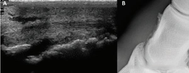
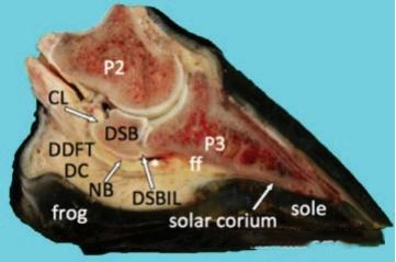
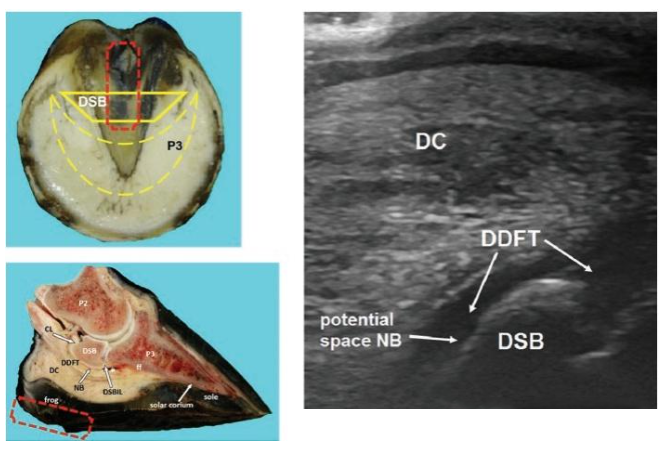
The hoof area contains crucial soft tissue structures highly sensitive to high-intensity exercise, including the distal portion of the deep digital flexor tendon (DDFT), the navicular bursa, collateral ligaments, and joint capsules. Ultrasound provides clear assessment of these structures.
Section II: Key Anatomical Focus for Ultrasound Examination
Ultrasound inspection of the equine hoof and pastern primarily covers the following structures vital to athletic performance:
| Key Structures | Common Injury Presentation |
| Distal Deep Digital Flexor Tendon (DDFT) | The most common site of injury in the hoof area. Ultrasound reveals tendon fiber continuity, potential disorganization, and the presence of hypoechoic lesions. |
| Collateral Ligaments of the DIP Joint | Highly susceptible to strain injury during high-intensity exercise. Imaging may show edema, fiber interruption, or localized enlargement. |
| Navicular Bursa (NB) | Bursitis is typically characterized by increased fluid within the sac and thickening of the wall. Ultrasound clearly demonstrates the bursa’s condition. |
| DIP Joint and Joint Capsule | Used to evaluate increased synovial fluid (effusion), capsular distension (suggesting arthritis or chronic strain), and inflammatory changes. |
Technical Note: Comprehensive visualization of these complex structures often requires the veterinarian to combine different planes (longitudinal/transverse) and various probe angles.
Section III: Interpreting Lesion Characteristics
Accurate diagnosis requires establishing a “baseline image” of healthy horses:
Normal Appearance: In a longitudinal view, the DDFT should appear as a highly organized, high-echoic band of parallel fibers. In a transverse view, tendons and ligaments should have a round cross-section and uniform internal echogenicity. Bursae and joint capsules should have clear walls and minimal fluid.
Pathological Signals: Any areas of hypoechoic darkening , fiber disruption or irregularity , an increase in cross-sectional area , or capsular distension are clear indicators of pathology.
Summary of Common Pathological Features:
| Injury Type | Key Ultrasound Features |
| Deep Digital Flexor Tendon Injury ,Hypoechoic dark area | Interruption of fiber alignment |
| Ligamentitis or Rupture ,Ligament thickening | Uneven echogenicity |
| Navicular Bursitis ,Increased fluid within the bursal cavity | Significant wall thickening |
| Joint Capsule Lesions ,”Increased echogenicity of synovial fluid | Capsular distension, indicating arthritis or chronic strain.” |
Section IV: Practical Application and Value
Hoof and pastern ultrasound holds broad significance throughout the athletic life of a horse:
Pre-Race Screening: Prevents horses with occult injuries from competing, thereby reducing the risk of catastrophic failure during the event.
Post-Competition Follow-up: Monitors for potential micro-trauma following high-intensity exercise.
Rehabilitation Guidance: Dynamic imaging assesses the progress of healing, allowing veterinarians to scientifically determine the optimal time for return to training.
Protecting Breeding Value: For high-value stallions/mares, early diagnosis of hoof issues can prevent the decline of their breeding utility.
The health of the hoof and pastern is paramount to the athletic longevity and performance of a sport horse. Ultrasound provides an unprecedented visualization tool, allowing veterinarians to literally “see” minute changes in tendons, ligaments, and bursae, facilitating effective early management.
Post time: Nov-11-2025