


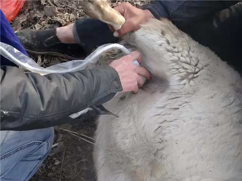
The area between the right side of the rib cage and the caudal edge of the ipsilateral scapula was shaved and alcohol solution was used to remove surface grease and dirt before performing ultrasound in sheep. A microconvex high-frequency probe (6-10 MHz) was used to perform the examination with the sheep in a standing position.A complete ultrasound examination of the liver was performed starting from the right side of the ribs and proceeding from the twelfth to the fifth intercostal space (IS).Each examination was performed from dorsal to ventral using both longitudinal and transverse complete scans.
The scanning area was divided into three different scanning windows: zone 1 (Z1) , from the right side of the ribs to the 11th IS; zone 2 (Z2) from the 10th IS to the 8th IS; and zone 3 (Z3) from the 7th IS to the 5th IS.The liver was also examined, with the probe placed in the cranial portion of the right side of the ribs (HYP). All detected encapsulated lesions were always localized and characterized according to WHO guidelines.
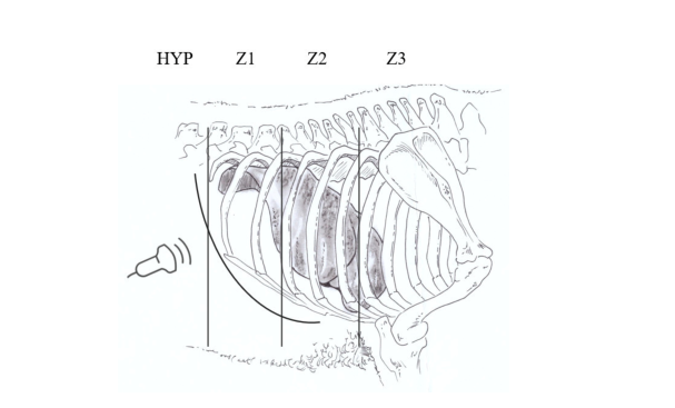
In the above figure: division of the liver region used to develop a rapid scanning procedure. hYP: liver scan by suspicion; Z1: liver scan from the ribcage to the 11th intercostal space; Z2: liver scan from the 10th to the 8th intercostal space; Z3: liver scan from the 7th to the 5th intercostal space. cl: unicompartmental anechoic cystic lesion with homogeneous anechoic content. The cyst wall is not visible.
CE1: homogeneous anechoic cyst with uniform anechoic content or fine internal echoes.
CE2: cyst with internal segregation, sometimes honeycomb-like.
CE3: uni-atrial cyst that may show the presence of a sub-cyst.
CE4: hypoechoic and hypoechoic matrix similar in appearance to a ball of wool.
CE5: cyst with partially or completely calcified cyst wall.
CE6: cystic cysts that have been detected by the ultrasound machine in the liver.
CE7: cysts that have been detected by the ultrasound machine in the liver.
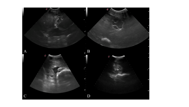
Liver lesions detected by a sheep ultrasound machine: (B) unicompartmental cysts with detachment of the lamina propria from the cyst wall, which may contain subcysts, transitional; (C) thick calcified walls that are arched, producing conical shadows and inactivity; and (D) liver abscesses.
Dawei sheep ultrasound machine for sale, if you want to know more about the price of ultrasound machine for sheep, please contact us, we will provide the best ultrasound machine for sheep
Post time: Apr-10-2024