


When a cow looks unwell, time matters. So does accuracy. And in the real world of bovine practice, those two pressures often collide in a muddy yard, a crowded chute, or a remote farm where decisions cannot wait for perfect conditions.
That is exactly why the question “cattle x ray vs ultrasound” keeps coming up.
Both tools are valuable. Both can change a case. But they do not answer the same diagnostic question. Not really. X ray and ultrasound overlap in a few areas, yet their strengths are fundamentally different. One is usually better for bone, mineralized structures, and certain thoracic views. The other shines in soft tissue, fluid-filled structures, reproductive work, and many chute-side examinations.
If you want the short answer, here it is:
For cattle, ultrasound is usually the first-choice imaging tool for reproduction and many soft-tissue or abdominal problems, while X ray is more useful when you need to evaluate fractures, bony lesions, mineralized changes, or selected thoracic and limb conditions.
Still, that simple summary hides the nuance. And nuance matters.
This guide breaks down the real differences between cattle X ray and ultrasound, explains where each method performs best, and shows how portable systems such as the RV-550A digital radiography unit and the Y6 veterinary ultrasound can fit into modern bovine workflows .
Quick Comparison: Cattle X Ray vs Ultrasound
| Factor | Cattle X Ray | Cattle Ultrasound |
| Best for | Bone, joints, mineralized lesions, foreign bodies with radiopacity, selected thoracic imaging | Reproduction, pregnancy diagnosis, soft tissue, fluid, abdominal organs, some thoracic and musculoskeletal applications |
| Weakness | Less informative for many soft-tissue problems without contrast; positioning and motion can be limiting | Gas and bone interfere strongly; image quality depends heavily on operator skill and acoustic access |
| Portability | Portable DR systems make field imaging more practical, but positioning and radiation control still matter | Typically easier for chute-side and on-farm use; highly practical in reproductive and soft-tissue work |
| Speed in field use | Fast image acquisition with digital systems, but setup and safety protocols are essential | Often faster for screening live, standing cattle in real farm settings |
| Radiation exposure | Yes | No ionizing radiation |
| Common bovine uses | Orthopedic checks, distal limb issues, trauma workups, some thoracic assessment | Pregnancy diagnosis, fetal aging, reproductive tract evaluation, abdominal screening, pleural/lung checks, soft tissue assessment |
What Is Cattle X Ray Used For?
Cattle X ray, or bovine radiography, uses ionizing radiation to create a projection image of internal structures. In plain language, it helps clinicians see differences in tissue density: gas, fat, soft tissue or fluid, bone or mineral, and metal or contrast material. That density-based contrast is exactly why radiography remains so relevant.
It is especially useful when the clinical question is structural.
Think fractures. Think claw or distal limb problems. Think bony proliferation, joint change, osteomyelitis, severe trauma, or a radiopaque foreign object. In those cases, X ray often gives a more direct answer than ultrasound ever could.
According to the Merck Veterinary Manual, radiography is a core diagnostic method for evaluating transmitted X-ray patterns and is particularly useful in musculoskeletal work and thoracic imaging, though correct positioning and motion control are critical for diagnostic quality. Merck also notes that as subject thickness increases, scatter becomes a greater problem, which is one reason large-animal radiography requires good technique and realistic case selection.
So no, X ray is not obsolete in cattle practice. Far from it. It simply needs to be used for the right problem.
Common cattle cases where X ray may help
- · Suspected fractures or fissures
- · Lameness involving distal limbs or joints
- · Hoof and claw-related bony changes
- · Trauma assessment
- · Evaluation of mineralized lesions
- · Some thoracic cases where radiographic density differences can be informative
In valuable breeding stock, performance animals, or referral-level cases, that information can be decisive.
What Is Cattle Ultrasound Used For?
Ultrasound works differently. Instead of X rays, it uses sound waves. That means no ionizing radiation, real-time imaging, and a completely different set of strengths.
In cattle, ultrasound is often the more practical everyday imaging tool. Not because it is universally better, but because bovine medicine asks many questions that ultrasound is particularly good at answering.
Reproduction is the obvious example. It is hard to overstate how important ultrasound has become in modern cattle management. It is used for pregnancy diagnosis, fetal aging, reproductive tract assessment, ovarian evaluation, and management decisions tied to breeding efficiency.
A University of Florida IFAS Extension publication notes that conventional B-mode ultrasound can detect a viable embryo at around 28 days after mating, while Doppler approaches can assess corpus luteum function even earlier in some reproductive programs. The same source highlights the role of ultrasound in fetal aging, reproductive scoring, resynchronization planning, and herd-level management decisions.
But reproduction is only part of the story.
The Merck Veterinary Manual describes veterinary ultrasonography as particularly useful for soft tissues in the abdomen, thorax, and musculoskeletal system, and for guiding procedures such as biopsies. It also points out the limits: gas reflects sound strongly, bone blocks the beam, and image interpretation depends a lot on operator experience.
That balance is the key to understanding ultrasound in cattle. It is excellent. But it is not magic.
Common cattle cases where ultrasound may help
- · Pregnancy diagnosis
- · Ovarian and uterine evaluation
- · Fetal viability and fetal age estimation
- · Abdominal organ assessment
- · Urinary or reproductive tract checks
- · Pleural or selected lung surface evaluation
- · Some tendon, ligament, or soft-tissue lesions
- · Procedure guidance in selected settings
Cattle X Ray vs Ultrasound: The Real Clinical Difference
Here is the simplest way to think about it:
- · X ray shows density differences best
- · Ultrasound shows soft tissue interfaces and fluid best
That sounds neat on paper. In practice, it means:
Choose X ray when you suspect bone or mineralized pathology
If the lesion lives in bone, involves a joint margin, or depends on visualizing mineral density, radiography is usually the stronger tool. It gives a broad structural overview and can reveal changes ultrasound may only hint at indirectly.
Choose ultrasound when you need real-time soft tissue information
If the case involves pregnancy, uterus, ovaries, abdominal viscera, pleural fluid, or a superficial soft-tissue structure, ultrasound often wins on speed, practicality, and diagnostic yield.
Choose both when the case is layered
This is where experienced clinicians tend to land.
A PubMed-indexed review on bovine orthopedics argued that both radiology and ultrasonography can improve the chance of reaching a definitive diagnosis, especially in valuable cattle. That is a useful reminder: the question is not always which one is better? Sometimes the right answer is which one first, and what should follow?
Is Ultrasound Better Than X Ray for Cattle?
Sometimes yes. Sometimes absolutely not.
If the problem is reproductive, abdominal, or soft tissue in nature, ultrasound is often the better first-line tool. It is fast, repeatable, practical in the field, and does not involve radiation. In many bovine settings, that combination is hard to beat.
If the problem is skeletal, heavily mineralized, or related to a fracture pattern, X ray may be the more decisive imaging method.
So the better question is not:
Which modality is better overall?
It is:
Which modality is better for this specific lesion, this specific animal, in this specific setting?
That shift in thinking prevents wasted time, blurred images, and expensive guesswork.
Field Reality Matters More Than Theory
A textbook answer is useful. A farm-ready answer is better.
In bovine practice, equipment choice is not just about image quality. It is about whether the system can actually be used where cattle live, move, and get treated. If an imaging unit is too cumbersome, too slow to deploy, or too fragile for field conditions, even strong specs on paper may translate into mediocre real-world value.
That is why portability has become a serious part of the imaging conversation.
Where a portable DR system fits: RV-550A
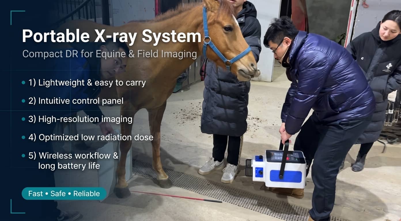
For bovine teams that need X ray capability beyond a fixed room, a portable digital radiography setup can make radiographic work far more realistic. The RV-550A is designed around that field-friendly logic.
Based on the product material provided, the RV-550A offers:
- · Portable DR design for mobile use
- · 5.6 kW output power
- · 40–125 kV exposure range
- · Wireless 17 × 17 inch flat panel detector
- · 139 μm pixel size and 16-bit ADC for detailed images
- · Preview time around 1.4 seconds and imaging time around 3.7 seconds
- · Built-in high-capacity battery for use where power access is limited
- · Wireless image transfer
- · Tripod-based foldable structure for transport and setup
- · Image processing software with DICOM connectivity
In practical terms, that makes the RV-550A relevant for bovine users who need veterinarydigital X ray performance in mobile or semi-mobile scenarios, especially where limb evaluation, trauma workup, or selective on-site radiography can improve decision-making.
Not every cattle case needs radiography. But when radiography is the right modality, mobility matters.
Where a handheld ultrasound fits: Y6

If your daily workload leans toward herd checks, reproductive work, abdominal screening, or fast soft-tissue evaluation, a compact ultrasound system may offer more day-to-day utility. The Y6 cattle ultrasound machine is built with that use pattern in mind.
From the official product information and related product listings, Y6 highlights include:
- · Handheld design for bovine and equine field use
- · About 1.35 kg weight
- · Fully waterproof body, washable after farm use
- · 8-inch HD display
- · 6600 mAh battery
- · B, BB, 4B, BM, and M imaging modes
- · 32 GB storage, expandable via TF card
- · Preset packages for multiple species, including cows
- · Abdomen, heart, obstetric, and superficial measurement functions
- · Optional field accessories such as display hood or goggle-style viewing for bright outdoor environments
That combination is especially attractive in cattle practice because it aligns with how ultrasound is often used: quickly, repeatedly, and sometimes under less-than-perfect conditions.
Small. Rugged. Washable. Ready to move.
That is not fluff. On a farm, those details matter.
When to Use Cattle X Ray Instead of Ultrasound
Use X ray first when:
1. You suspect a fracture or bone lesion
Ultrasound may show surrounding soft-tissue changes, but it will not replace radiographic evaluation of many bony problems.
2. You need a density-based view
Mineralized structures, some foreign bodies, and osseous change are better matched to radiography.
3. You are assessing selected limb and joint problems
Especially where structural alignment or cortical integrity matters.
4. You need digital image documentation for referral or follow-up
A modern portable DR unit can support this workflow efficiently.
When to Use Cattle Ultrasound Instead of X Ray
Use ultrasound first when:
1. The question is reproductive
Pregnancy diagnosis, ovarian structures, uterine status, fetal viability, fetal age. This is classic ultrasound territory.
2. You suspect a soft-tissue or fluid-related problem
Ultrasound is often much more revealing for these cases.
3. You need a fast chute-side answer
Especially in herd management, screening, or repeated examinations.
4. Radiation logistics would slow the case down
Ultrasound is often easier to deploy in everyday farm conditions.
Can X Ray and Ultrasound Be Used Together in Cattle?
Yes, and often they should be.
This is not a contradiction. It is good medicine.
A lame cow with swelling near a joint may need ultrasound to assess adjacent soft tissue and X ray to characterize osseous change. A traumatic case may benefit from ultrasound as a rapid screening tool in one stage and radiography as a confirmatory structural tool in the next. A herd veterinarian may rely on ultrasound every day, then bring in DR selectively for cases where skeletal detail changes the treatment plan.
The smartest imaging workflow is rarely ideological. It is sequential.
How to Choose the Right Imaging Tool for Bovine Practice
If you are deciding what to add to your clinic, truck, or farm service workflow, ask these five questions:
1. What kind of cases do you see most?
- · Mostly reproduction and herd checks? Ultrasound will likely bring more daily value.
- · More trauma, lameness, and structural diagnostics? DR may deserve priority.
2. How often do you work in the field?
The more mobile your practice is, the more important durability, battery life, setup speed, and transportability become.
3. Do you need radiation-free routine imaging?
If yes, ultrasound has obvious practical advantages.
4. Do you need better skeletal detail?
If yes, X ray remains essential.
5. Are you building a complementary imaging workflow rather than a single-tool workflow?
That is often the best long-term answer. One tool does not erase the need for the other.
Final Verdict: Cattle X Ray vs Ultrasound
So, cattle X ray vs ultrasound — which one wins?
Neither wins outright.
Ultrasound is usually the more versatile everyday tool for bovine reproductive and soft-tissue work. X ray is the more decisive tool for fractures, bony pathology, and selected structural cases.
If your goal is practical, repeatable, field-based diagnosis, ultrasound often becomes the frontline option. If your goal is to visualize mineralized anatomy with clarity, radiography still carries the edge.
And in a modern bovine practice, the strongest setup may not be choosing one over the other. It may be knowing exactly when to deploy each.
That is where products like the Y6 and RV-550A make sense. The Y6 aligns with fast, portable, high-frequency bovine scanning workflows. The RV-550A supports digital radiography where skeletal detail, image documentation, and mobile DR capability matter. Different jobs. Different strengths. Same clinical objective: a better answer, faster.
Frequently Asked Questions
Is ultrasound or X ray better for pregnancy diagnosis in cattle?
Ultrasound is better. It is widely used for pregnancy diagnosis, fetal viability assessment, reproductive tract evaluation, and fetal aging in cattle.
Can an X ray detect pregnancy in cattle?
It is not the preferred tool. In bovine practice, pregnancy diagnosis is typically performed with palpation, ultrasound, or other reproductive management methods rather than routine radiography.
What is the best imaging tool for cattle lameness?
It depends on the cause. If the issue is primarily bony or involves suspected fracture, X ray may be more useful. If soft tissue involvement is suspected, ultrasound can add important information.
Is portable DR practical for cattle?
Yes, in selected cases. Portable DR systems can make bovine radiography more accessible in field or semi-field settings, especially for trauma and orthopedic work, though radiation safety and positioning remain essential.
Why is handheld ultrasound popular in cattle practice?
Because it is fast, mobile, repeatable, and highly useful for reproductive and soft-tissue examinations. In herd settings, those advantages are hard to ignore.
Post time: May-11-2026