


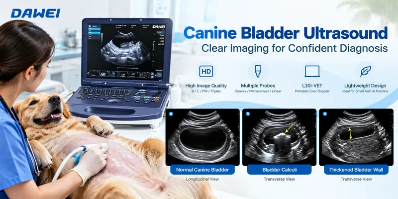
Canine bladder ultrasound is an important imaging technique in small animal veterinary practice. It allows veterinarians to assess the urinary bladder’s shape, wall thickness, internal contents, and surrounding structures without using ionizing radiation.
Whether examining a dog with urinary difficulties or performing a routine abdominal examination, selecting the appropriate ultrasound probe and understanding the resulting images are essential for a thorough assessment.
Quick Answer: For canine bladder ultrasound, a microconvex probe is a practical choice for routine examinations, particularly in small and medium-sized dogs. A high-frequency linear probe offers better detail of superficial bladder walls, while a lower-frequency convex8 probe may be more appropriate for deeper examinations in large dogs. The optimal choice depends on body size, bladder depth, and the clinical question.
This guide explains probe selection, scanning techniques, common ultrasound findings, and the application of the Dawei L30i-VET portable color Doppler ultrasound system in canine urinary examinations.
1. Why Is Bladder Ultrasound Important for Dogs?
The urinary bladder is responsible for storing urine before it leaves the body. Changes in its wall, internal contents, or surrounding structures may be associated with urinary tract disorders.
Veterinarians commonly perform bladder ultrasound when dogs present with symptoms such as:
- Frequent or difficult urination
- Blood in the urine (hematuria)
- Recurrent urinary tract infections
- Suspected urinary bladder stones
- Abdominal discomfort
- Suspected bladder masses or abnormalities
Unlike conventional radiography, ultrasound provides direct visualization of fluid-filled structures and surrounding soft tissues.
For example, a veterinarian may observe echogenic material within the bladder, evaluate a suspected wall abnormality, or investigate the presence of urinary calculi.
However, ultrasound findings alone cannot always determine the underlying cause. Urinalysis, urine culture, radiography, and other diagnostic procedures may be necessary.
2. Which Ultrasound Probe Is Best for Canine Bladder Examination?
Probe selection is one of the most important factors influencing image quality.
A suitable probe should provide sufficient imaging depth while maintaining the resolution needed to visualize the bladder wall and contents.
Microconvex Probe: A Versatile Choice
Microconvex probes feature a relatively small contact surface and a curved imaging field.
They are particularly useful when examining small dogs or scanning through narrow acoustic windows.
Advantages include:
- Compact probe footprint
- Convenient access to the caudal abdomen
- A widening field of view with depth
- Practical visualization of the bladder and surrounding structures
For routine examinations, a microconvex probe operating at an appropriate frequency can provide a useful balance between penetration and anatomical detail.
Linear Probe: Better Superficial Detail
High-frequency linear probes are valuable when the bladder is located close to the abdominal wall.
They provide higher spatial resolution at shallow depths, making them useful for evaluating:
- Bladder wall thickness
- Wall contour and layering
- Small superficial structures
- Selected focal abnormalities
Their main limitation is reduced penetration at higher operating frequencies.
Convex Probe: Useful for Larger Dogs
In larger or deep-bodied dogs, the bladder may be located farther from the skin surface.
A lower-frequency convex probe can provide greater penetration and a wider imaging field.
This makes it useful for evaluating deeper abdominal structures, although very fine superficial detail may be less apparent than with a high-frequency linear probe.
Probe Comparison for Canine Bladder Ultrasound
| Probe Type | Main Advantage | Typical Application |
|---|---|---|
| Microconvex | Small footprint and versatile depth | Routine examination in small and medium dogs |
| Linear | High-resolution superficial imaging | Bladder wall and superficial detail |
| Convex | Greater penetration | Deeper imaging in larger dogs |
Clinical Tip: Select the highest practical frequency that still provides adequate penetration to the target anatomy. Probe geometry, patient body condition, bladder filling, and acoustic access should all influence the final choice.
3. How to Perform a Canine Bladder Ultrasound
A systematic scanning approach helps ensure that important anatomical regions are not overlooked.
Step 1: Prepare the Patient
The dog may be positioned in dorsal or lateral recumbency, depending on its condition and the examination requirements.
The caudal abdomen is prepared to ensure effective contact between the ultrasound probe and the skin.
Ultrasound gel is applied to reduce air interference and improve acoustic transmission.
For a complete bladder examination, moderate bladder filling is generally preferable. An insufficiently filled bladder can make wall evaluation more difficult.
Step 2: Locate the Urinary Bladder
Position the ultrasound probe on the ventral caudal abdomen, just cranial to the pelvic region.
The urinary bladder typically appears as a fluid-filled structure with predominantly dark internal contents.
Adjust the imaging depth until the bladder and its surrounding boundaries are clearly visible.
Step 3: Obtain Longitudinal and Transverse Images
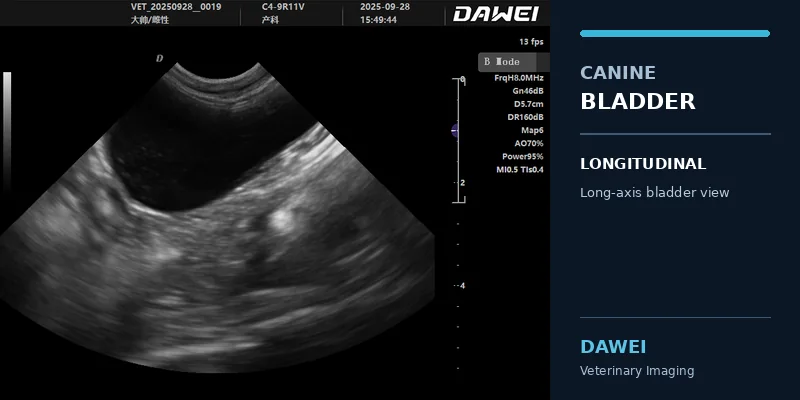
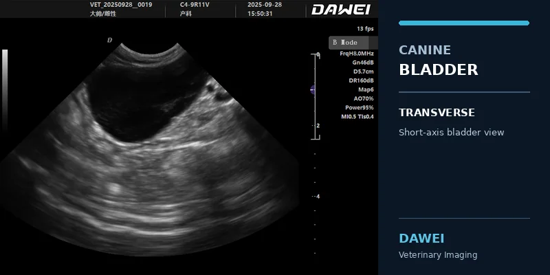
A complete evaluation should include both longitudinal and transverse imaging planes.
In the longitudinal plane, assess the bladder from its cranial apex toward the bladder neck.
In the transverse plane, scan systematically through the same regions while observing the wall contour and internal contents.
Particular attention should be paid to the bladder trigone and proximal urethra when visible.
Step 4: Optimize the Ultrasound Image
Important adjustments include:
Depth: Display the entire bladder with enough surrounding anatomy for orientation.
Gain: Adjust overall brightness while preserving the appearance of fluid-filled structures.
Focus: Position the focal zone near the bladder wall or structure being assessed.
Frequency: Balance spatial resolution and tissue penetration according to the dog’s size.
Time Gain Compensation (TGC): Adjust brightness at different depths when necessary.
These settings can help reduce image interpretation errors caused by poor visualization.
Step 5: Document the Findings
Record images showing the bladder in both scanning planes.
If a focal abnormality is identified, document its location, appearance, dimensions, and relationship to the bladder wall.
When wall thickness is measured, account for bladder filling and use an appropriate measurement plane.
A complete urinary tract examination should also consider the kidneys, visible ureters, and urethra according to the clinical indication.
4. What Does a Normal Dog Bladder Look Like on Ultrasound?
A normally filled canine urinary bladder generally appears as a rounded or oval fluid-containing structure.
On B-mode ultrasound, urine is typically anechoic, meaning that it appears black because it produces very few internal echoes.
The bladder wall normally has a relatively smooth contour.
Its apparent thickness depends partly on the degree of bladder distension. A relatively empty bladder may have a thicker or irregular-looking wall without necessarily indicating disease.
Image Example 1: Canine Bladder Ultrasound – B-Mode Imaging
[Insert actual L30i-VET canine bladder ultrasound image: longitudinal view]
Figure 1. Longitudinal B-mode ultrasound view of a canine urinary bladder. The image should demonstrate the fluid-filled bladder lumen and surrounding wall. Image acquired using Dawei L30i-VET; anatomical interpretation should be verified by a qualified veterinarian before publication.
This type of image helps veterinarians assess bladder shape, lumen appearance, and wall continuity.
A transverse view should also be obtained to provide additional anatomical information.
5. Common Abnormal Findings in Canine Bladder Ultrasound
Ultrasound can reveal structural changes that require further clinical evaluation.
Understanding how these findings appear is important for veterinary practitioners.
5.1 Urinary Bladder Stones
Bladder stones, also called uroliths, may appear as bright echogenic structures within the bladder lumen.
Larger or sufficiently mineralized structures often produce posterior acoustic shadowing, visible as a dark region extending beyond the object.
However, small calculi may not produce a clearly detectable acoustic shadow.
Changing the dog’s position may help determine whether the echogenic material moves within the bladder.
Image interpretation: Bright intraluminal structures with acoustic shadowing may raise suspicion of bladder stones, but the examination should be correlated with other clinical findings.
Image Example 2: Suspected Bladder Calculi
[Insert an authentic, veterinarian-confirmed canine bladder calculus ultrasound image if available]
Figure 2. Example of echogenic material within the canine urinary bladder. If a confirmed calculus image is available, identify the calculus and associated acoustic shadow. Do not label an unverified image as bladder stones.
5.2 Bladder Wall Thickening
Bladder wall thickening may be associated with inflammatory changes, chronic urinary disorders, or other abnormalities.
However, apparent thickening can also occur when the bladder is insufficiently distended.
The veterinarian should consider bladder volume, patient size, clinical symptoms, and any additional findings before interpreting wall thickness.
Ultrasound alone cannot reliably distinguish bacterial cystitis from sterile inflammation.
5.3 Urinary Sediment
Echogenic particles may sometimes be visible within the bladder lumen.
These findings may represent urinary sediment, cellular material, crystals, or other contents.
Importantly, image artifacts can mimic sediment.
Adjusting the probe angle and evaluating whether the material moves with gravity can help clarify the appearance.
Urinalysis may be needed to determine the nature and significance of the material.
5.4 Bladder Masses and Focal Lesions
Focal soft tissue structures may occasionally be detected along the urinary bladder wall.
Their ultrasound appearance can vary in shape, echogenicity, and vascularity.
Color Doppler imaging may help evaluate vascular signals associated with a suspected wall lesion.
Nevertheless, ultrasound imaging cannot independently establish whether a mass is inflammatory, benign, or malignant.
Further testing may be necessary.
6. Can Color Doppler Help During Bladder Examination?
B-mode ultrasound is generally the primary imaging method for evaluating the shape, wall, and contents of the canine urinary bladder.
Color Doppler provides additional information when vascular assessment is relevant.
For example, Doppler imaging may help investigate blood-flow signals in a suspected soft tissue lesion or distinguish certain vascular structures from surrounding tissue.
However, Doppler findings must be interpreted carefully. The absence or presence of visible flow alone does not establish a definitive diagnosis.
Image Example 3: Color Doppler Examination
[Insert actual L30i-VET canine bladder color Doppler image only if available and clinically relevant]
Figure 3. Canine urinary bladder assessment using B-mode and color Doppler imaging. Color Doppler may provide supplementary vascular information when clinically indicated.
7. Dawei L30i-VET for Canine Bladder Ultrasound
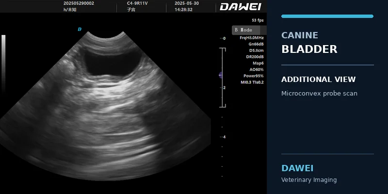
For veterinary clinics performing routine abdominal and urinary examinations, the choice of ultrasound system can affect both imaging flexibility and workflow.
The Dawei L30i-VET is a portable, laptop-style veterinary color Doppler ultrasound system designed for small animal examination and other veterinary applications.
It supports abdominal imaging, urinary system assessment, reproductive examinations, and selected vascular applications when configured with appropriate probes.
L30i-VET Probe Options for Bladder Imaging
| Probe | Frequency Range | Practical Use |
|---|---|---|
| R11 Microconvex | 4.0–8.0 MHz | Routine small-animal abdominal and bladder examinations |
| Linear | 6.0–12.0 MHz | Superficial bladder wall detail |
| Convex | 2.0–5.5 MHz | Deeper abdominal examinations in larger dogs |
The standard configuration includes the R11 microconvex probe. Other compatible probes can be selected according to clinical requirements.
Additional Imaging and Workflow Features
The L30i-VET offers:
- At least a 15-inch display
- B-mode and color Doppler imaging
- PW spectral Doppler and real-time triplex imaging
- Tissue harmonic imaging and spatial compound imaging
- Adjustable gain, depth, focal settings, and image parameters
- Examination presets and measurement functions
- DICOM 3.0 support and image storage options
These features support imaging acquisition, examination review, and clinical documentation.
For canine bladder examinations, correct probe selection and image optimization remain more important than using advanced Doppler modes in every case.
Explore the Dawei L30i-VET Veterinary Ultrasound System
8. Frequently Asked Questions About Dog Bladder Ultrasound
What is the best probe for a dog bladder ultrasound?
A microconvex probe is often a practical starting choice for general canine bladder imaging. A high-frequency linear probe may provide better superficial detail, while a convex probe may be needed for greater penetration in larger dogs.
Can ultrasound detect bladder stones in dogs?
Ultrasound can identify many bladder stones as echogenic structures, sometimes with posterior acoustic shadowing. However, very small stones and sediment can be difficult to distinguish, and supplementary tests may be needed.
Can a veterinarian diagnose cystitis using ultrasound alone?
No. Ultrasound may reveal bladder wall thickening or other changes compatible with cystitis, but the cause cannot be established from imaging alone. Clinical examination, urinalysis, and urine culture may be necessary.
Does a dog’s bladder need to be full for ultrasound?
Moderate bladder filling generally improves visualization of the bladder wall and internal contents. Both underfilling and excessive distension can affect interpretation.
Can the Dawei L30i-VET be used for canine urinary examinations?
Yes. The L30i-VET supports small-animal abdominal and urinary imaging with compatible microconvex, linear, or convex probes. The appropriate probe and settings depend on the dog’s anatomy and examination requirements.
9. Conclusion
Canine bladder ultrasound is a valuable component of small animal diagnostic imaging.
Successful examinations depend on appropriate probe selection, suitable bladder filling, systematic scanning in multiple planes, and careful interpretation of ultrasound findings.
Microconvex probes are versatile for routine urinary examinations, high-frequency linear probes offer additional superficial detail, and convex probes can provide greater depth when needed.
For veterinary clinics seeking a portable imaging system, the Dawei L30i-VET combines multiple probe options, B-mode imaging, color Doppler capabilities, and clinical workflow features in a laptop-style platform.
Choosing equipment according to patient size and examination requirements is essential for obtaining useful diagnostic images.
Looking for a Veterinary Ultrasound Machine for Your Clinic?
Dawei Veterinary Medical provides veterinary ultrasound solutions for pet hospitals, veterinary clinics, and animal care professionals.
Contact our team to discuss your clinical applications, probe requirements, and suitable equipment configurations.
Clinical References
- Seiler GS, et al. ACVR and ECVDI consensus statement for the standardization of the abdominal ultrasound examination. Veterinary Radiology & Ultrasound. 2022. https://doi.org/10.1111/vru.13151
- Pease A. Ultrasonographic Evaluation of the Urinary Bladder in Dogs & Cats. Clinician’s Brief. Updated July 2024. https://www.cliniciansbrief.com/article/ultrasound-bladder-animals-cystitis-urinary
- Dawei Veterinary Medical. L30i-VET Veterinary Color Doppler Ultrasound Technical Specifications. https://www.dwvet.com/pet-ultrasound-machine-for-sale-veterinary-doppler-system-product/
Post time: Oct-29-2026