


Although several methods have been reported for pregnancy diagnosis in sheep, ultrasound, especially B/W ultrasound, is considered to be the best choice for pregnancy diagnosis due to its high resolution, real-time performance and accuracy. In a sheep farm where pregnant sheep are separated from nulliparous sheep and managed in separate flocks, B/W ultrasound is used to separate pregnant from nulliparous sheep.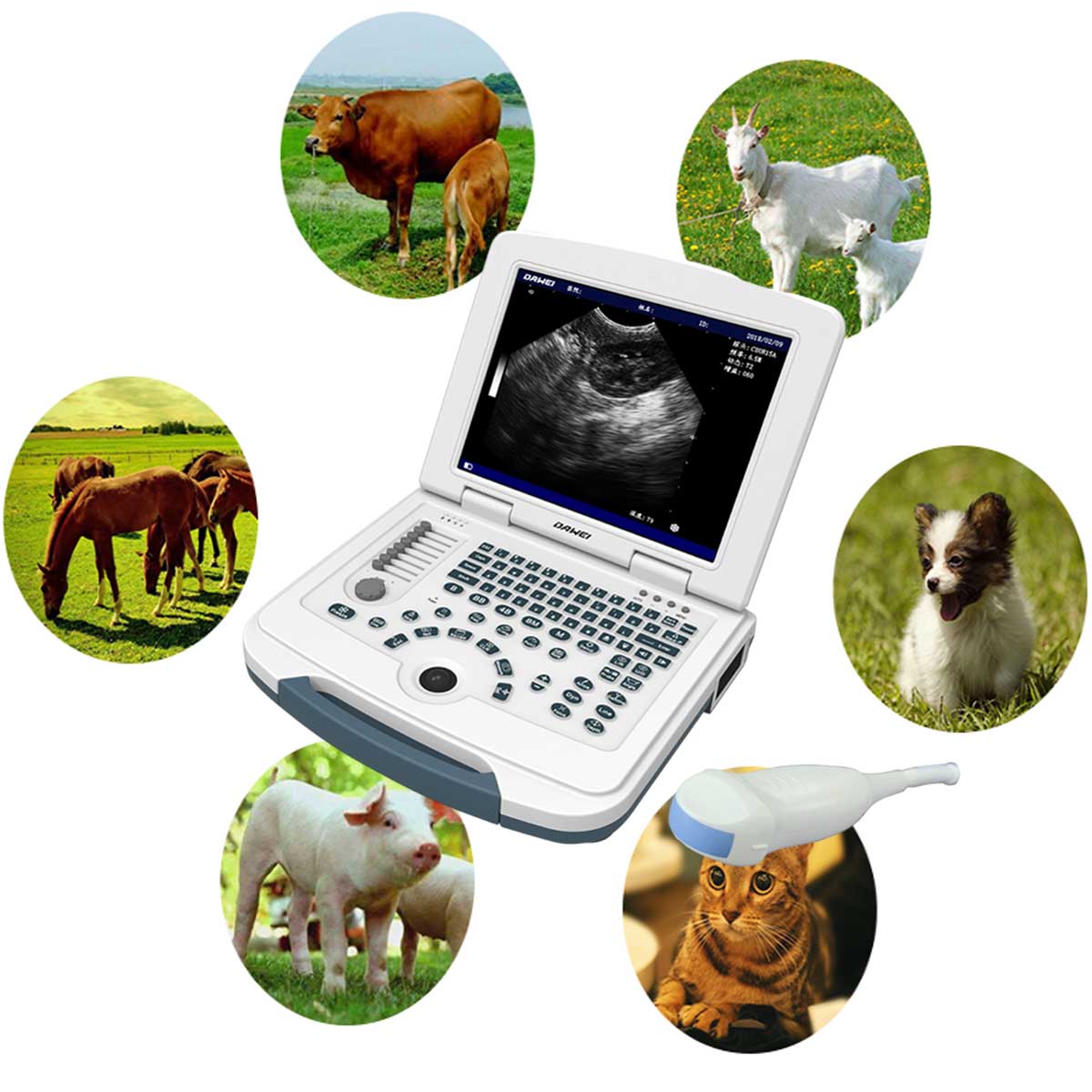
1 Examination equipment
The MU10 fully digital portable vet ultrasound machine system manufactured by Dawei Veterinary Medical.
2 Examination method
The sheep stood in the pen and the examiner was positioned at the back of the sheep’s side. First, the ultrasonic diagnostic instrument display was adjusted to the maximum brightness, and then the control panel was adjusted to adjust the depth , total gain , and probe frequency according to the actual display.
The probe was coated with coupling agent, the examiner controlled the back of the sheep with the left hand, placed the probe in the groin of the hind limb with the right hand, and slid it forward along the base of the atrium and swept down the side of the abdomen, terminating at the mid-abdomen to form a complete “7″ shape examination.
In addition, when ultrasound is performed in an open barn, ambient light interferes with the visualization of the image display, so it is important to increase the brightness of the image to minimize ambient light interference. First, select as much of the backlit area as possible prior to the examination; then adjust the screen brightness to maximum, adjust the total gain until the sidewalls of the probe are clearly visible, and finally apply a couplant to the probe to reduce air and light interference. In addition, placing a piece of cardboard on top of the screen as a light shield also helps to display the screen image.
3 Examination results
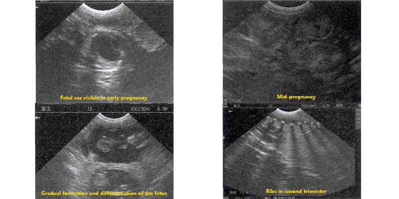
3.1 Sonographic characteristics of the pregnant uterus
In early pregnancy, the image shows an echo-less round or oval sac with a moderately echogenic long oval fetal body, see Figure 1. In the middle stage of pregnancy, there is a large liquid dark area in the uterus, and in the dark area, there are several “copper-coin-like” uterine lobules, see Figure 1. The fetus is mostly located in the lower abdomen, and it is gradually shaping up, and the structures such as cephalic, trunk and caudal side can be differentiated, see Figure 1. The fetus is located in the lower abdomen and is gradually taking shape, with differentiation of the cephalic, trunk, and caudal structures, as seen in Figure 1. The image of the fetus is clearer in the second trimester, with obvious fluctuations in the fetal heart and the structure of the fetal heart, as well as the complete calcification of the bones of the spine and limbs, as seen in Figure 1.
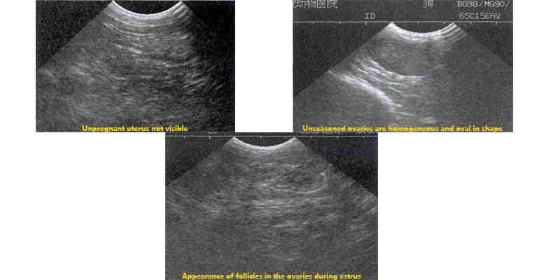
3.2 Sonographic features of the non-pregnant uterus
The normal non-pregnant uterus is seen behind the base of the udder and appears as a moderately echogenic mass of uniform texture surrounded by a peritoneal membrane, see Fig. 2. The ovarian structures are sometimes visible when examined cephalad along the uterus. The ovaries of anestrous sheep appear as homogeneous isoechoic oval structures, see Fig. 2, while those of anestrous sheep may be irregular in shape, with hypoechoic to anechoic nodules in the parenchyma, see Fig. 2.
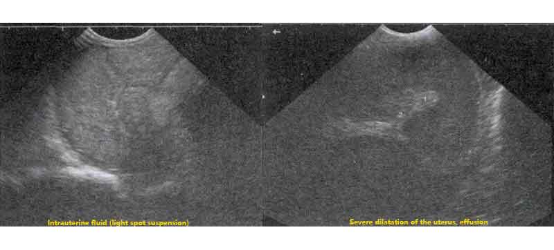
3.3 Sonographic features of the diseased uterus
During the examination, it was found that some of the uteri of the non-pregnant sheep showed pathologic conditions, the most common of which was uterine effusion. The fluid-filled uterus was usually dilated and elongated, with a flow of fluid containing a large number of suspended light spots, see Fig. 3. In severe cases of fluid-filled uterus, the cavity of the uterus was dilated and formed a mid-gestation uterus, with a large number of irregular liquid dark areas, but no uterine lobules or fetal morphology were observed, see Fig. 3.
Ultrasound examination of pregnant sheep is not only fast, simple and accurate, but also has the ability to differentiate the gestation cycle, monitor fetal status and show uterine lesions, which is a good guide for practical work.
Post time: Nov-23-2023