


In the fast-paced world of veterinary medicine, diagnostic imaging is the linchpin of effective treatment. Whether you are running a busy 24-hour emergency hospital or a specialized referral center, the choice between animal digital radiography (DR) and veterinary ultrasound determines the speed and accuracy of your diagnosis. While both modalities are non-invasive cornerstones of animal X-ray imaging, they answer fundamentally different physiological questions.
Leading providers of veterinary solutions, such as Dawei Veterinary Medical, emphasize that a complete diagnostic suite integrates veterinary DR, veterinary ultrasound, and veterinary ECG monitoring into a seamless ecosystem. This guide explores the clinical logic behind choosing the right modality for every patient—from a 500g exotic pet to a 100kg Mastiff—ensuring your practice delivers the gold standard in care.
1.The Core Distinction: Physics Meets Physiology
The decision matrix begins with tissue density. Veterinary digital X-ray systems utilize ionizing contrast structures like bone and air-filled organs. In contrast, ultrasound uses high-frequency radiation to create a 2D projection map of density, making them the superior choice for high sound waves to interpret acoustic impedance, excelling at visualizing the internal architecture of soft tissues and fluid dynamics.
Choose DR For:
- Skeletal Integrity: Fractures, luxations, osteoarthritis, and spinal dysplasia.
- Thoracic Evaluation: Lung patterns (alveolar, interstitial), Heart size (VHS), and tracheal collapse.
- Abdominal Survey: Radio-opaque foreign bodies, GDV (bloat), and organ size overview.
- Dental: Periodontal disease, root abscesses, and jaw integrity.
Choose Ultrasound For:
- Soft Tissue Architecture: Liver texture, kidney cortex definition, adrenal glands.
- Fluid Dynamics: Ascites, pleural effusion, and blood flow velocity (Doppler).
- Real-Time Function: Cardiac contractility (Echocardiography), intestinal peristalsis.
- Reproduction: Fetal viability (heartbeats) and early pregnancy confirmation.
2.Clinical Scenarios: Canine and Feline Patients
In daily practice, dogs and cats present with symptoms that often require a “DR first” approach for rapid screening. For a coughing Cavalier King Charles Spaniel, a thoracic DR image is indispensable for calculating the Vertebral Heart Score (VHS) and assessing pulmonary edema. The high resolution of modern detectors-such as those with 3.6 LP/mm spatial resolution-allows clinicians to distinguish between bronchial and interstitial patterns with clarity. For gastrointestinal issues, such as a vomiting Labrador, canine DR provides an immediate overview. It can instantly identify radio-opaque foreign bodies (like stones or toys) or characteristic gas patterns indicating obstruction. If the X-ray is inconclusive, ultrasound becomes the logical next step to evaluate bowel wall thickness or identify intussusceptions. This tiered approach maximizes diagnostic yield while managing client costs.
3.Special Considerations: Exotics and Small Mammals
Imaging a 500g guinea pig or a parakeet presents unique challenges compared to a Great Dane. These patients have rapid heart and respiratory rates, making motion blur a significant adversary. A premium veterinary DR system must offer extremely short exposure times-down to 1.Oms-to “freeze” motion effectively. Furthermore, “burning out” thin skeletal structures is a common risk. Systems with a wide dynamic range capable of low-energy settings (starting at 40kV) are essential. The use of high-sensitivity Cesium lodide (Csl) detectors allows for lower radiation doses, protecting these smaller, more radiation-sensitive patients while maintaining the high contrast needed to visualize minute bone fractures or dental pathology in rabbits.
4.Handling the Giants: Large Breed Requirements
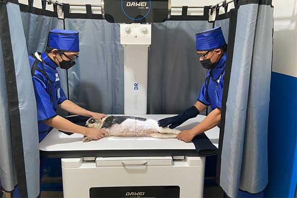
Conversely, large breeds like Mastiffs or St. Bernards require raw power and physical robustness. Penetrating a deep chest or a dense abdomen demands a generator capable of delivering high kV 400mA) is the industry standard for ensuring diagnostic quality in these heavy patients. and mAs without extending exposure time. A 32kW generator (delivering up to 150kV and Physical safety is equally important. A wobbly table can cause panic in a nervous giant breed. Veterinary-specific tables, like those found in the Dawei RV-32B series, feature a four-way floating design with a load capacity of 2100kg, allowing staff to position heavy, sedated animals safely and precisely without moving the patient manually.
5.The Integrated Workflow: DR, Ultrasound, and ECG
The most efficient clinics do not view these tools in isolation. A “same-visit” workflow integrates them for a comprehensive picture. For a cardiac case, the workflow typically begins with veterinary DR to assess overall heart size and lung fluid. This is followed by veterinary ultrasound (Echocardiography) to measure contractility and valve function. Finally, veterinary EcG provides the electrical data needed to diagnose arrhythmias. Seamless connectivity is the glue of this workflow. Systems supporting GigE data interfaces ensure that large, high-resolution X-ray images (often 3072×3072 pixels) are transferred to the PACS workstation in s1 second. This allows the veterinarian to show owners the “whole picture” in one exam room, improving compliance and treatment outcomes.
6.Summary Comparison: DR vs. Ultrasound
| Feature | Digital Radiography (DR) | Ultrasound |
| Primary Indication | Bones, Lungs, Trauma Survey, Dentistry | Soft Tissue, Organ Architecture, Pregnancy |
| Image Physics | Density Projection (2D) | Acoustic Impedance (Cross-sectional) |
| Speed | Rapid Triage (Preview ≤1s) | Procedure Dependent (Minutes to Hours) |
| Radiation Control | Low Dose (CsI Scintillator Efficiency) | None (Non-ionizing) |
| Field of View | Large (e.g., 430mm × 430mm) | Narrow (Probe footprint) |
| Durability | Liquid Resistant (IPX1) & High Load | Delicate Probes (Drop sensitive) |
| Connectivity | High Speed (GigE / Wireless) | DICOM / USB Export |
A Unified Diagnostic Front In modern veterinary practice, the question is rarely “which one?” but “which one first?” Veterinary DR provides the roadmap, while veterinary ultrasound acts as the magnifying glass. By investing in reliable, high-specification equipment—from powerful 32kW generators to high-definition probes —clinics position themselves to handle any case with confidence. Manufacturers like Dawei Veterinary Medical support this mission by delivering robust, integrated imaging ecosystems designed for the realities of animal care.
Frequently Asked Questions
Q: Can Digital Radiography detect pregnancy in pets?
A: Yes, but only after the fetal skeletons calcify, which occurs around day 42-45 of gestation. For earlier detection (day 20-25) and viability checks (heartbeats), veterinary ultrasound is the superior tool.
Q: Why is a Cesium Iodide (CsI) detector better for animals?
A: CsI scintillators are more efficient at converting X-rays into light than older materials. This means you can get a high-quality image with a significantly lower radiation dose—crucial for smaller animals and staff safety.
Q: How does generator power (kW) affect image quality in large dogs?
A: A higher power generator (e.g., 32kW) allows for shorter exposure times even at high dosage levels. This “freezes” the image, preventing motion blur caused by panting or trembling in large patients.
Q: What are the installation requirements for a veterinary DR system?
A: While older systems required 3-phase power, modern units like the Dawei RV-32B are designed for standard single-phase 220VAC power, greatly simplifying installation in existing clinic spaces.
Q: Is the equipment waterproof?
A: Veterinary environments are prone to accidents. Look for detectors with at least an IPX1 rating, which protects against vertical dripping water and urine, ensuring longevity.
Q: How quickly can I see the X-ray image?
A: Efficiency is key. High-quality systems with GigE connectivity provide an image preview in ≤1 second, allowing for immediate assessment and reducing the time an animal needs to be restrained.
Post time: Jan-13-2026