


Quick Answer
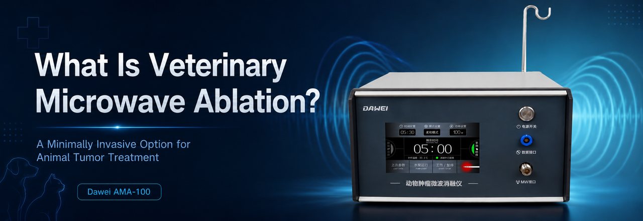
Veterinary microwave ablation is a local thermal treatment in which a veterinarian places a thin microwave antenna into or near a tumor and delivers non-ionizing electromagnetic energy to heat and destroy targeted tissue. Placement is usually guided by ultrasound, computed tomography (CT), fluoroscopy, laparoscopic ultrasound, or direct surgical visualization. Depending on the tumor and access route, the treatment may be performed through a small skin puncture, during laparoscopy, or as an adjunct to open surgery.
The technique has been reported most often in dogs with selected liver tumors, small pulmonary nodules, retroperitoneal masses, and bone lesions. It has also been used for palliative control of bleeding from ulcerated mammary tumors. However, the published veterinary evidence remains limited and largely case-based; microwave ablation is not a universal replacement for surgery, chemotherapy, radiation therapy, or other oncologic care.
In one sentence: Microwave ablation for animals uses an image-guided antenna to create a controlled zone of heat inside a selected tumor while limiting treatment of surrounding tissue.
What Is Veterinary Microwave Ablation?
Veterinary microwave ablation—often shortened to MWA—is a form of animal tumor ablation. Instead of removing a mass, the procedure aims to destroy targeted tumor tissue in place.
A veterinarian inserts a narrow ablation antenna into the lesion. The system then delivers microwave-frequency electromagnetic energy through the antenna. That energy creates rapid molecular motion within the tissue, producing heat through dielectric heating. When the target tissue reaches a sufficient temperature for an adequate period, proteins denature, cell membranes fail, and a zone of coagulative necrosis forms.
That definition sounds straightforward. The difficult part is precision.
The treatment team must plan the antenna path, place the active tip correctly, create an ablation zone large enough to cover the intended target, and protect critical structures nearby. Imaging before, during, and after treatment is therefore central to the procedure—not an optional extra.
How Does Microwave Ablation Work in Animals?
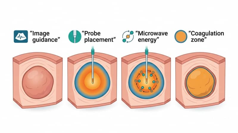
1. The tumor is identified and mapped
The veterinary team uses diagnostic imaging to assess the tumor’s location, size, shape, depth, relationship to vessels or organs, and whether a safe access path exists. Ultrasound and CT are commonly reported guidance tools in canine studies.
2. The microwave antenna is placed
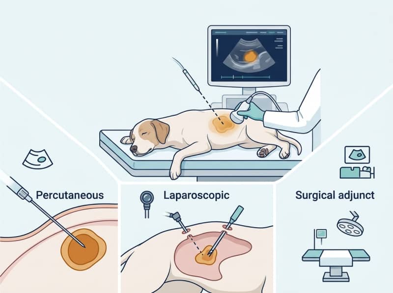
Under appropriate anesthesia and sterile conditions, a veterinarian advances an ablation antenna into the planned position. The approach may be percutaneous, laparoscopic, or performed during surgery. One antenna position may be sufficient for a small target; other lesions may require overlapping treatment zones.
3. Microwave energy creates local heat
An oscillating electromagnetic field causes polar molecules—especially water molecules—to rapidly reorient. This molecular movement produces heat within the target tissue. Unlike surgical excision, the tumor is not physically removed during the ablation itself.
4. Heat produces a coagulation zone
Sustained thermal exposure damages cells and denatures proteins. The intended result is a defined zone of nonviable tissue that encompasses the target, often with a planned safety margin when anatomy allows. Follow-up imaging is used to assess the treated area and watch for residual or recurrent disease.
Why Is It Considered Minimally Invasive?
Microwave ablation is often described as minimally invasive because it can sometimes reach an internal tumor through a small skin puncture rather than a large incision. In reported canine cases, percutaneous procedures have been guided by ultrasound or CT, and some patients were discharged the same day or within a few days.
But “minimally invasive” does not mean noninvasive, painless, or risk-free.
The antenna still enters the body. Accurate imaging, anesthesia or sedation, sterile technique, pain control, thermal planning, and post-procedure monitoring are required. Some tumors cannot be safely reached percutaneously. In those cases, a laparoscopic or open surgical approach may give the clinician better visibility and control.
Common access routes
| Access route | What it means | Potential use |
| Percutaneous | The antenna passes through a small skin puncture under image guidance | Selected internal tumors with a safe, visible access path |
| Laparoscopic | A camera and instruments enter through small abdominal ports | Lesions that benefit from direct visualization or laparoscopic ultrasound |
| Open surgical adjunct | MWA is performed during an operation | Tumors found to be unresectable, multifocal, or difficult to remove completely |
The least invasive route is not automatically the safest route. Selection depends on anatomy, tumor biology, operator experience, available imaging, and the animal’s overall condition.
Which Animal Tumors May Be Considered?
Published veterinary reports do not establish a single, universally accepted list of indications. They do show where clinicians have explored the technology—primarily in dogs.
Liver tumors
The liver has the largest published veterinary MWA experience to date, although the numbers remain small.
A 2017 case series described five dogs with primary or metastatic hepatic neoplasia. All had diffuse disease that prevented complete surgical resection; lesions measuring 0.5 to 2.5 cm were treated during laparotomy without procedural complications. The authors described MWA as feasible and potentially useful as an adjunct in appropriately selected dogs with nonresectable hepatic tumors.
A separate four-dog series reported percutaneous, ultrasound-guided MWA for three primary and one metastatic hepatocellular carcinoma. Procedures lasted 10 to 40 minutes at 30 to 35 W, with no immediate complications reported. Follow-up ranged from 39 to 649 days, and the authors recommended regular imaging plus larger studies before effectiveness could be established.
A 2024 case report also described laparoscopic ultrasound-guided ablation of a canine hepatocellular carcinoma. The treated mass showed complete ablation on CT at three and 12 months, while a separate liver lesion later enlarged and required resection. This distinction matters: successful local treatment of one lesion does not prevent new or separate disease elsewhere.
Retroperitoneal and renal-region masses
In a three-dog report, two renal masses and one retroperitoneal mass near the diaphragm were treated percutaneously using ultrasound and CT guidance. The masses measured 1.4 to 2.5 cm. No intraprocedural or major postprocedural complications were reported, and imaging findings after treatment were consistent with successful local treatment.
These results are encouraging, but three cases are not enough to define broad safety or survival benefit.
Solitary pulmonary nodules
A preliminary prospective study described percutaneous MWA of solitary presumed pulmonary metastases in two dogs previously treated for appendicular osteosarcoma. Ultrasound or CT guidance was used. Pneumothorax occurred in both dogs and required treatment in one; survival after MWA was 82 days in one dog and 19 days in the other.
The authors concluded that the procedure was technically feasible, while emphasizing that additional studies were needed to determine survival benefit and confirm tumor-cell death. Accordingly, lung ablation should be presented as a highly selected, specialist procedure—not routine treatment.
Bone lesions
A pilot study evaluated MWA in six dogs with distal radial osteosarcoma. Histopathology showed variable tumor necrosis, ranging from 30% to 90%, with a median of 55%. No immediate intraoperative or periprocedural complications were observed.
The study was designed to test feasibility and safety, not to prove durable local control or replace amputation. Later single-case reports have combined MWA with cementoplasty as part of multimodal limb-sparing care, but complications such as pathological fracture remain possible.
Bleeding, ulcerated mammary tumors
MWA has also been reported as a palliative hemostatic technique. In two elderly dogs with bleeding ulcerated mammary tumors, treatment under local anesthesia achieved complete hemostasis and subsequent scarring of the ulcerated site.
This use case is different from curative-intent tumor ablation. The immediate objective was to stop bleeding and improve comfort when conventional surgery or general anesthesia was problematic.
What Makes a Patient a Possible Candidate?
A veterinary oncologist or interventional clinician may consider MWA when several conditions align:
- · The disease is localized enough for a defined ablation target.
- · Imaging shows a feasible and reasonably safe antenna path.
- · The planned thermal zone can be created without unacceptable injury to nearby organs, vessels, nerves, skin, bowel, diaphragm, or other critical structures.
- · Surgery is not possible, would be unusually extensive, or needs a local adjunct.
- · The procedure has a clear goal: local control, cytoreduction, symptom relief, or palliation.
- · The animal can tolerate the required anesthesia or sedation and post-procedure monitoring.
- · The owner understands that local ablation does not necessarily treat microscopic, multifocal, or metastatic disease.
Tumor type alone is not enough. Size, shape, location, margins, tissue characteristics, blood supply, prior treatment, metastatic status, and the clinician’s equipment and experience all influence suitability.
Potential Benefits
For a carefully selected animal, potential advantages may include:
- · Small access site: Percutaneous treatment may avoid a large incision.
- · Focused local therapy: Energy is delivered at the tumor rather than throughout the body.
- · Flexible procedural routes: MWA can be percutaneous, laparoscopic, or used during surgery.
- · Real-time image guidance: Ultrasound, CT, fluoroscopy, or laparoscopic ultrasound can support placement and monitoring.
- · Possible treatment of unresectable targets: Some reported canine liver tumors were treated when complete surgical removal was not possible.
- · Repeatability in selected cases: A new or residual local target may sometimes be reassessed for another image-guided procedure, although this depends on anatomy and clinical judgment.
- · Palliative applications: The technique may help control a specific symptom, such as bleeding from an ulcerated mass.
These are potential procedural advantages, not guarantees of tumor control or longer survival.
Risks and Limitations
The principal risk of any thermal ablation is damage outside the intended treatment zone. Possible complications depend heavily on the organ and access route and may include:
- · bleeding or hematoma;
- · burns or unintended thermal injury;
- · injury to bowel, diaphragm, skin, nerves, vessels, ducts, or adjacent organs;
- · infection;
- · pneumothorax when treating lung lesions;
- · incomplete ablation, residual viable tumor, or local recurrence;
- · needle-track complications;
- · anesthesia-related events;
- · post-ablation pain, inflammation, fever, or malaise.
The evidence base is another major limitation. The canine publications cited here include case reports, small case series, and one pilot study with six dogs. They support technical feasibility in selected settings, but they do not establish that veterinary MWA improves survival across tumor types.
Most importantly, animal tumor ablation is local therapy. It cannot be assumed to control disease that has already spread or to replace systemic treatment when chemotherapy, immunotherapy, targeted therapy, or other care is indicated.
Veterinary Microwave Ablation vs. Other Treatments
| Treatment | Primary action | Key distinction |
| Surgical excision | Removes the tumor and, when possible, surrounding margins | Provides a surgical specimen and may remain preferred for resectable disease |
| Microwave ablation | Heats and destroys tissue in situ | May reach selected lesions through a small access route; no intact tumor specimen is removed |
| Radiofrequency ablation | Uses alternating electrical current to generate heat | Different energy delivery; comparative veterinary evidence is limited |
| Cryoablation | Freezes tissue to induce cell injury and death | Creates an ice ball that may be visible on imaging; availability varies |
| Radiation therapy | Uses ionizing radiation delivered over one or more sessions | Does not require antenna placement inside the tumor |
| Chemotherapy or systemic therapy | Treats disease throughout the body | Addresses systemic risk but may not provide the same immediate local destruction |
These modalities are not mutually exclusive. MWA may be considered within a multimodal plan rather than as a stand-alone replacement for every other therapy.
What Happens Before, During, and After Treatment?
Before treatment
The team confirms the diagnosis when feasible, stages the disease, reviews bloodwork and coagulation status, evaluates anesthesia risk, and plans the antenna path with imaging. The treatment goal should be explicit: complete local ablation, reduction of tumor burden, control of bleeding, pain relief, or another defined endpoint.
During treatment
The patient is positioned and monitored. Imaging guides antenna placement. The clinician selects treatment settings and may reposition the antenna to create overlapping zones. Temperature monitoring or protective techniques may be used when vulnerable structures are nearby.
After treatment
Immediate imaging can look for expected ablation changes and complications. The patient receives analgesia and monitoring appropriate to the organ treated. Scheduled follow-up imaging is essential because an ablation zone evolves over time and because residual, recurrent, or new disease may not be clinically obvious.
Frequently Asked Questions
Is veterinary microwave ablation the same as surgery?
No. Surgery removes tissue; MWA destroys targeted tissue in place using heat. MWA can be delivered percutaneously, laparoscopically, or during an open operation, so it may complement surgery rather than replace it.
Is microwave ablation for animals noninvasive?
No. It is more accurately described as minimally invasive when performed through a small puncture or laparoscopic access. An antenna still enters the body, and anesthesia, imaging, sterile technique, and monitoring are usually required.
Does MWA use radiation?
MWA uses non-ionizing electromagnetic energy to generate heat. It is not the same as ionizing radiation therapy. CT may be used for guidance in some procedures, while others use ultrasound.
Which species can receive veterinary microwave ablation?
Most published clinical evidence identified for this article involves dogs. A specialist may evaluate other species, but evidence from canine reports should not be generalized automatically to cats, horses, or exotic animals.
Can veterinary microwave ablation cure cancer?
It may achieve local destruction of a selected lesion, but “cure” depends on tumor type, stage, margins, microscopic disease, metastatic risk, and follow-up. Current veterinary evidence is too limited to promise cure or a survival advantage across cancers.
Can the treatment replace chemotherapy?
Not necessarily. MWA is local; chemotherapy and other systemic therapies address disease beyond the treated site. Some patients may need both, while others may not be candidates for either. The treatment plan must be individualized.
How is the ablation zone checked?
Clinicians may use ultrasound or CT immediately after treatment and at scheduled follow-up visits. Imaging helps evaluate coverage, treatment response, complications, and possible recurrence.
What should a pet owner ask the veterinary team?
Useful questions include: What is the exact treatment goal? Why is MWA preferred over surgery or radiation in this case? What imaging will guide the procedure? Which structures are at risk? How will pain be managed? What follow-up is required? What evidence supports MWA for this specific tumor?
Veterinary microwave ablation is a targeted, image-guided thermal therapy that may offer a minimally invasive route to local tumor treatment in carefully selected animals. Published canine reports support feasibility in liver, lung, retroperitoneal, bone, and palliative mammary-tumor applications. Still, the evidence is early and heterogeneous. The most responsible use of MWA is therefore case-specific, specialist-led, and integrated with diagnosis, staging, risk assessment, and long-term imaging follow-up.
Post time: Sep-04-2026