


Choosing a veterinary color doppler ultrasound system is not only a purchasing decision. It is a clinical decision, an operational decision, and, in many practices, a long-term strategic decision.
That distinction matters.
A lower-cost scanner may appear attractive on paper. A feature-heavy platform may look impressive in a demo. Yet neither is automatically the right choice. In veterinary imaging, the systems that create lasting value are the ones that support accurate interpretation, efficient workflow, repeatable image acquisition, and a realistic fit with the clinic’s caseload.
In other words, what matters most is not how many functions a machine claims to have. It is how reliably those functions contribute to diagnosis.
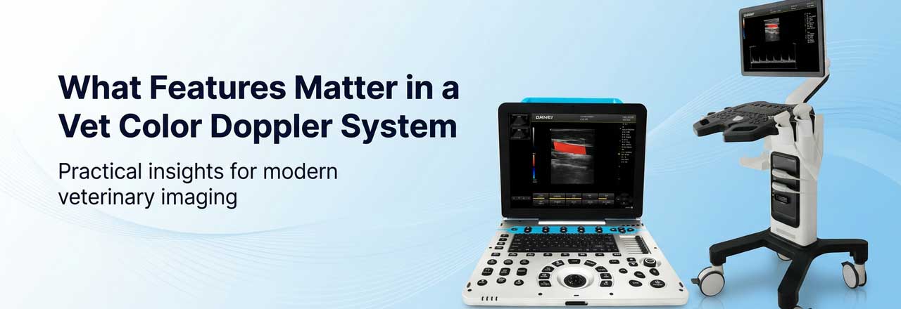
This article explains the core features that matter in a vet Color Doppler system, why they matter clinically, and how buyers can assess them more critically. It also shows how different system tiers can be positioned naturally within a practical veterinary imaging portfolio, with light-touch brand alignment to Dawei Veterinary Medical.
Why Color Doppler matters in veterinary imaging
Ultrasound is widely valued in veterinary medicine because it helps clinicians evaluate soft tissues in real time, especially abdominal organs. In cardiac work, Doppler ultrasound adds another layer of diagnostic relevance by helping the examiner evaluate blood flow rather than structure alone.
That is the key shift.
A grayscale image may show anatomy. A Doppler-enabled exam can help the clinician understand hemodynamics.
Veterinary references consistently describe color and spectral Doppler as important for evaluating blood-flow direction, blood-flow velocity, and turbulent flow patterns. Merck Veterinary Manual also notes that echocardiography is excellent for confirming tentative diagnoses, assessing the severity of valvular regurgitation and stenotic lesions, detecting chamber enlargement, identifying congenital defects, and evaluating cardiac structure and function with the support of 2D, M-mode, color, and spectral Doppler imaging.
This is why a true veterinary color doppler system should be evaluated as a diagnostic platform, not just a machine with colored flow display.
1. Foundational B-mode image quality is still the first priority
Many buyers are drawn to Doppler terminology because it sounds advanced. However, the clinical value of Doppler depends heavily on the quality of the underlying 2D image.
If B-mode imaging is weak, then vessel borders may be unclear, lesion margins may be poorly defined, and color overlays may become visually impressive but diagnostically limited. A reliable animal sonography machine therefore needs strong grayscale performance before Doppler functions are even considered.
Important image-quality features include:
- · Tissue harmonic imaging
- · Speckle or noise reduction
- · Spatial or compound imaging
- · Adjustable dynamic range
- · Multiple focal zones
- · Fine gain and TGC control
- · Adequate penetration without losing too much detail
Why do these functions matter? Because veterinary scanning is rarely performed under ideal conditions. Small patients move. Large dogs have deep abdomens. Cats may present with subtle parenchymal changes. Reproductive and emergency cases often allow very little time for trial-and-error optimization.
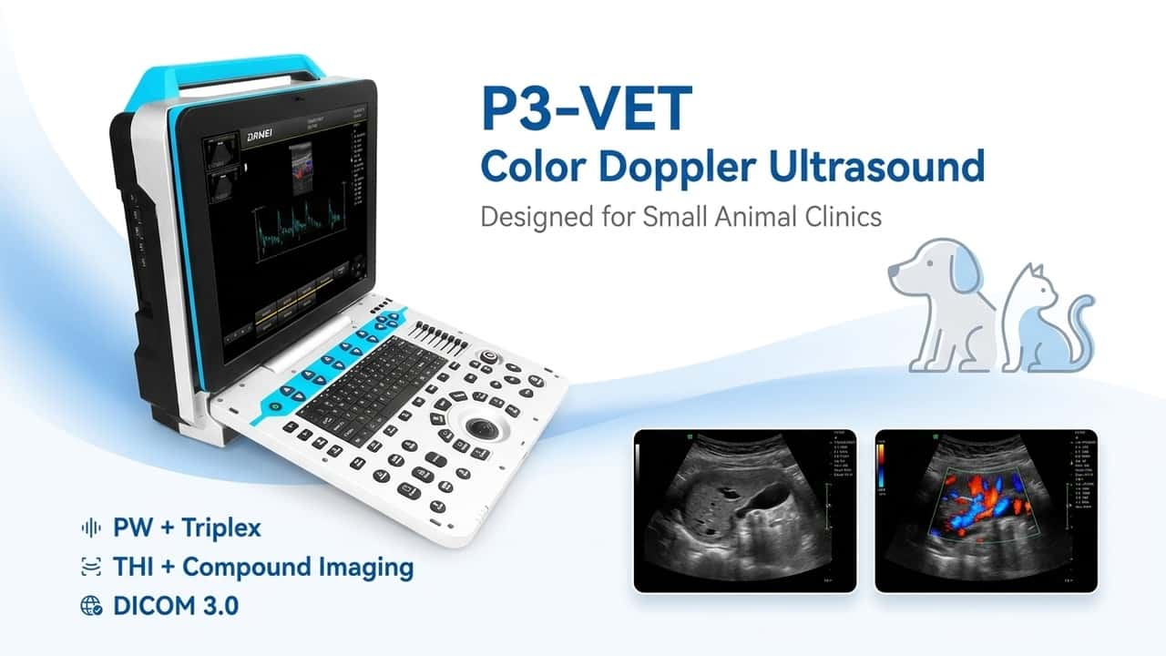
Within the Dawei Veterinary Medical -Veterinary Ultrasound portfolio, clinics can find systems that combine Tissue Harmonic Imaging (THI), compound imaging, trapezoidal imaging, and workflow-friendly optimization tools. For example, models such as the P3-VET and F3-VET represent the kind of practical Color Doppler platforms many clinics consider when they want a capable pet ultrasound machine rather than a system that only sounds advanced in marketing language.
2. A complete Doppler package matters more than color display alone
When people say they want Color Doppler, they often mean they want better cardiovascular insight. But color flow display is only one part of that workflow.
Color Doppler helps visualize the direction and general behavior of blood flow on top of the grayscale image. It is useful for locating abnormal jets, recognizing flow disturbance, and quickly surveying regions of interest. Still, in many cases, the real diagnostic value appears when spectral Doppler is added.
For that reason, buyers should look beyond whether a machine has color. They should ask whether the system provides a meaningful Doppler package, including:
- · PW (Pulsed Wave Doppler) for site-specific velocity sampling
- · CW (Continuous Wave Doppler) for higher-velocity flow assessment
- · Power Doppler or directional power Doppler for low-flow sensitivity
- · Triplex imaging for simultaneous B-mode, color, and Doppler review
- · Adjustable sample volume
- · Angle correction
- · Spectral tracing or auto measurement tools
- · Clear velocity scale and baseline control
This is especially relevant for a canine ultrasound machine used in practices with internal medicine or cardiology demand. Color helps the operator see where flow is abnormal. Spectral Doppler helps quantify what that abnormality means.
This distinction can also be seen across the Dawei Veterinary Medical product range, where different system tiers offer different levels of Doppler capability—from practical everyday Color Doppler workflows to more advanced packages that include broader spectral and tissue Doppler support. In a cardiac-capable workflow, those are not decorative features. They are the difference between visual detection and deeper hemodynamic assessment.
3. Probe ecosystem is one of the most important purchasing criteria
A high-spec console cannot compensate for an incomplete or poorly matched probe strategy.
Veterinary imaging is inherently variable. One clinic may focus on small-animal abdominal studies. Another may need routine echocardiography. Another may serve mixed practice, reproduction work, superficial soft tissue cases, and occasional vascular assessment. Because of that, probe compatibility and probe range should be treated as central evaluation criteria.
In most veterinary settings, buyers should consider access to:
- · Micro-convex probes for versatile small-animal abdominal work
- · Convex probes for deeper penetration
- · Linear probes for superficial structures, vessels, and higher-resolution detail
- · Phased-array or cardiac probes for echocardiography
- · Rectal probes for reproduction-related applications where relevant
This is one of the easiest ways to assess whether a pet ultrasound machine is suited for real practice rather than demo-room performance.
Across the Dawei Veterinary Medical veterinary ultrasound lineup, probe flexibility is one of the more practical strengths. Depending on the system tier, buyers can access combinations of convex, linear, micro-convex, rectal, and cardiac-oriented probe options. For clinics with multi-doctor use, diversified case types, or plans to expand imaging services, this kind of flexibility can have direct operational value.
4. Cardiac capability should be assessed with discipline
Not every Color Doppler system should be positioned as a strong cardiology solution. This is where buyers—and marketers—need to be precise.
A machine may have color Doppler and still be a better fit for abdominal and general-purpose use than for regular echocardiography. If a clinic plans to scan cardiac patients routinely, it should examine whether the system supports the features needed for meaningful hemodynamic interpretation.
A more cardiology-ready veterinary color doppler system should ideally include:
- · A phased-array or dedicated cardiac probe option
- · PW and CW Doppler
- · Stable triplex display
- · Tissue Doppler options where applicable
- · Sufficient frame rate and image stability
- · Cardiac measurement and reporting workflow
- · Adjustable sampling volume, velocity scale, and baseline
This point is supported by authoritative veterinary references. Merck Veterinary Manual notes that echocardiography is useful for assessing valvular regurgitation, stenotic lesions, chamber enlargement, congenital disease, myocardial function, and pulmonary hypertension. Veterinary cardiology references also emphasize the importance of color and spectral Doppler in examining abnormal flow direction, velocity, and turbulence.
That is why cardiac claims should be made carefully. Within the Dawei Veterinary Medical portfolio, some systems are better suited to general companion-animal imaging, while higher-tier configurations—such as the P60-VET—may offer a better fit for buyers seeking stronger cardiology-oriented functionality.
5. Portability should be matched to real workflow, not treated as a lifestyle feature
Portability changes how often a machine gets used.
This is not a marketing phrase. It is a workflow fact.
A system that is easy to move between rooms may support more frequent use in general practice, emergency areas, treatment spaces, or bedside rechecks. A fixed cart-based platform may be more appropriate for high-volume imaging rooms, multi-probe workflows, or clinics that perform more formal and prolonged exams.
In practical terms, there are three broad use patterns:
- · Cart-based systems for heavier caseloads, multiple active probes, and advanced imaging workflows
- · Portable laptop-style systems for balanced mobility and capability
- · Handheld systems for screening, field use, or complementary quick checks
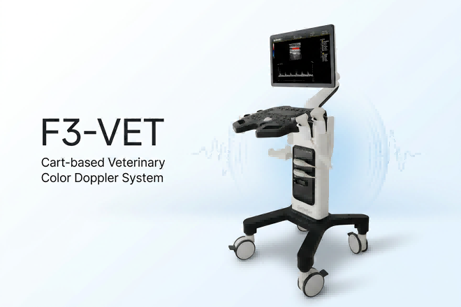
This same portfolio logic applies across the Dawei Veterinary Medical range. Some systems are designed for trolley-based workflows with larger displays and more probe connectivity. Others are built as portable or laptop-style animal sonography machine options for more flexible deployment. Handheld black-and-white units can also play a useful supporting role in rapid checks or field scenarios, but they should not be presented as substitutes for a full veterinary color doppler system.
6. Presets, optimization tools, and exam efficiency are clinically relevant
A machine should not demand excessive manual adjustment every time a new patient is scanned.
In busy veterinary settings, operators often work under time pressure. Patients may be anxious, unstable, or difficult to position. A system with stronger preset logic and optimization tools can reduce friction and improve consistency, especially when more than one doctor uses the scanner.
Key workflow-supporting features include:
- · Organ-specific or exam-specific presets
- · One-key image optimization
- · Easy parameter reset
- · Smooth cine review
- · Accessible measurement tools
- · Consistent clipboard and image-saving workflow
In the Dawei Veterinary Medical veterinary ultrasound range, workflow-oriented functions such as presettable inspection conditions, optimization tools, and user-friendly exam setup are exactly the kinds of details that matter in daily use—especially when positioning a canine ultrasound machine for general veterinary clinics rather than specialist-only environments.
7. Ergonomics and interface quality influence diagnostic consistency
Ergonomics are often discussed late in the buying process, even though they affect image acquisition every day.
Screen size, screen angle, interface responsiveness, image review flow, and measurement accessibility all shape the scanning experience. Over time, operator fatigue and workflow friction can influence consistency as much as raw imaging specifications do.
Across the Dawei Veterinary Medical veterinary ultrasound portfolio, screen size, interface layout, probe connectivity, and portability vary by system class. These are not the only factors that matter, but they deserve more attention than they usually receive in buying discussions.
8. Upgrade path and software headroom affect long-term ownership value
The best purchase is not always the machine that matches current demand exactly. Sometimes it is the one that supports realistic growth.
A clinic may begin with abdominal and reproductive studies, then gradually take on more cardiac work. A distributor may need a platform that can serve current market demand while leaving room for future differentiation. A hospital group may prefer systems that can be standardized but still scaled across use levels.
That is why upgradeability and software headroom matter.
Across the Dawei Veterinary Medical product range, some higher-tier systems also include broader functionality such as tissue Doppler, elastography, panoramic imaging, extended field-of-view, and 3D/4D-related features. Not every buyer needs these tools immediately. However, their presence can improve long-term product positioning and extend the useful life of the investment.
9. Species coverage and veterinary software support are practical differentiators
An effective animal sonography machine should support the kinds of patients a clinic actually sees.
That sounds elementary, but in practice it shapes usability. Species-specific presets, measurement packages, and veterinary workflow logic can reduce adjustment time and improve reporting consistency.
Across the Dawei Veterinary Medical veterinary ultrasound portfolio, species coverage and veterinary software support are important differentiators. In broader product-line positioning, these details are especially useful for SEO content intended to serve mixed-practice, breeding, or farm-animal search intent—not only companion-animal traffic.
10. The best system is the one that fits the clinic’s clinical reality
There is no universal best veterinary color doppler platform.
There is only the system that best matches the clinic’s case mix, operator profile, mobility needs, diagnostic expectations, and budget strategy.
A smaller companion-animal practice may prioritize a portable pet ultrasound machine with strong abdominal performance, straightforward Doppler, and a practical probe mix. A busier hospital may need a cart-based platform with more active interfaces, a larger screen, and stronger cardiac functionality. A distributor may need a complete product ladder, from handheld screening units to advanced Color Doppler systems, so that customers can upgrade without leaving the brand ecosystem.
This is where the Dawei Veterinary Medical veterinary ultrasound portfolio can be positioned naturally and credibly: with a product structure that spans portable systems, cart-based systems, and more advanced configurations for clinics with broader diagnostic needs. In practical content positioning, P3-VET can be framed as a portable everyday option, F3-VET as a cart-based workflow choice, and P60-VET as a more advanced system for clinics seeking broader functionality.
Final takeaway
When evaluating a veterinary color doppler system, the most important question is not whether the machine has color. It is whether the system helps clinicians generate reliable, interpretable, and repeatable diagnostic information across the cases they actually manage.
That requires a combination of:
- · strong B-mode image quality,
- · a meaningful Doppler package,
- · appropriate probe support,
- · workflow efficiency,
- · realistic cardiac capability,
- · and a product format that fits daily use.
For SEO purposes, this also creates a stronger article. It is more educational, more evidence-based, and more useful to both readers and search systems. For commercial purposes, it helps your product mentions feel natural instead of forced. And for AI retrieval, clear structure plus explicit clinical reasoning improves the chance that the article will be understood as a practical, trustworthy resource rather than a thin keyword page.
That is what stronger content looks like.
FAQ
Is Color Doppler always necessary for veterinary ultrasound?
No. For some routine screening tasks, a B/W scanner may be enough. However, clinics that perform cardiac work, vascular assessment, advanced internal medicine, or more detailed case evaluation often benefit from a veterinary color doppler system.
What is the most important feature in a pet ultrasound machine?
There is no single answer, but foundational B-mode image quality is often the first thing to evaluate. Without a strong grayscale image, Doppler functions will have limited clinical value.
What makes a canine ultrasound machine more suitable for cardiology?
A more cardiology-capable canine ultrasound machine typically includes a phased-array probe option, PW and CW Doppler, triplex imaging, stable frame performance, and measurement tools that support echocardiographic interpretation.
Can a portable animal sonography machine still be advanced?
Yes. Many portable platforms now offer Color Doppler, harmonic imaging, compound imaging, and advanced measurement functions. The key is balancing mobility with probe compatibility, image quality, and clinical workload.
Post time: May-13-2026