


Do you know how small animal ultrasound selects and manipulates probes when performing urinary tract, adrenal gland, and gastrointestinal tract exams?The following article gives you the answer。
1 Urinary tract and adrenal gland exploration techniques
1.1 Urinary tract
A 5.0 MHz probe is appropriate for most dogs, while a 7.5 MHz probe is used in cats. If necessary, higher frequency probes can be used for supplemental exploration of the left kidney in dogs and both kidneys in cats. The abdomen is clipped and a coupling agent is applied. The supine animal is examined for kidneys starting from the lower abdomen. It is easiest to locate the kidneys when in transverse section, whereas in sagittal section it is easy to do a complete examination of the kidneys. It is important to begin by pressing firmly on the probe to displace the overlying bowel loops and slowly sweep the entire kidney in each plane. The left kidney is easy to visualize. Because of its more posterior position, sometimes the spleen can provide an acoustic window for it. In the dog, the right kidney is more anteriorly positioned, within the costal arch, and is difficult to visualize because of the overlying inflated bowel loops. In cats, the kidneys are easier to explore due to their smaller size and more posterior location. Additional exploration is often necessary to adequately evaluate the kidneys. Scanning from the right 11 to 12 intercostal spaces yields high-quality images of the right kidney. The kidneys can be explored from the anterior lumbar paraspinal region while the animal is lying prone or standing, and this method of examination is particularly advantageous when the animal has a lot of ascites or a large mass because it reduces the distance of the probe from the kidneys. If the kidneys are not fully explored, some errors may occur when interpreting and illustrating the renal images; therefore, it is important to patiently explore the kidneys in multiple planes to ensure that high-quality results are obtained.
1.2 Adrenal glands

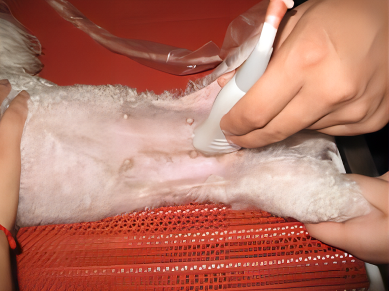
Adrenal gland exploration in dogs is usually performed with a 5.0 MHz or 7.5 MHz probe. The method is similar to renal exploration, with the animal lying supine, left lateral or right lateral, and scanning transversely, longitudinally or frontal from the lower or lateral abdomen. Hair must be removed to obtain a suitable adrenal visualization from the ventral side in most cases. Observations must avoid intestinal gas. In lateral recumbency, the superficial adrenal gland can be imaged or the lowermost portion of the adrenal gland can be imaged by probing with a Plexiglas table with a square hole in the center. The right adrenal gland is most easily explored from the right anterior abdomen at the 11th or 12th intercostal space. The left adrenal gland is most easily explored from the left side of the abdomen and occasionally from the 12th intercostal space. However, even with these techniques, the adrenal glands may not be visualized. Because of the small size of the adrenal glands, there is a risk of inconsistent visualization of the overlying inflated organs. The right adrenal gland is more difficult to visualize than the left.
1.3 Lower urinary tract
The lower urinary tract is commonly explored with a 7.5 MHz or 10.0 MHz probe, and a 5.0 MHz probe is used in large dogs when adjacent structures need to be evaluated. The use of a padding block assists in visualization of the lower bladder wall to minimize multiple reflection artifacts and to keep the bladder area within the focusing zone of the probe. If the bladder does not have a sufficient volume of urine, a small amount of diuretic can be injected or a bladder catheter can be used to introduce sterile saline to dilate the bladder. However, a general examination should be performed first. The bladder should be explored in two views from below the abdomen. If bladder wall thickness or a mass lesion is suspected, the probe should be oriented so that accurate measurements are obtained by passing the sound beam perpendicularly through the wall of the suspected area, which requires moving the probe laterally over the posterior abdomen. Partial thickening or multiple reflection artifacts may be seen occasionally as a result of intraluminal irritation or wall abnormalities, or may be due to the emission of the sound beam close to the bladder edge. Widening of the sound beam causes the lumen to echo laterally as if it were inside the bladder. Multiple reflected echoes caused by the probe, skin, or adjacent pneumatic bowel lumen can also be confused with intravesical echoes. These artifacts can be ruled out by shifting the position of the probe or by multiplanar imaging of the bladder. Different parts of the urethra in both females and males can be imaged from the lower abdomen. The urethra posterior to the prostate in males is usually not visible because it is obscured by the pubic bone or sciatic bone. Occasionally, the long axis of the urethra can be visualized from the perineum or by using a small line array probe with the rectum as the acoustic window. If necessary, the membranous urethra or penile urethra can be imaged using a padded block after removal of the coat. Very small specialized fan-scan probes or intracavitary rotary probes can be used to explore the bladder and urethra of the animal. Ultrasound endoscopic probes can also be used for transurethral exploration of the bladder.
2 Gastrointestinal tract exploration techniques
A real-time sector scanner with a probe frequency of 5.0 MHz or 7.5 MHz is used. For evaluation of the gastrointestinal tract wall layers, 7.5 MHz or a short-focus 5.0 MHz probe is recommended. Before ultrasound scanning the animal’s lower abdomen is clipped and a coupling agent is applied. The animal is often placed in a supine position, but in some cases it is placed in left or right lateral recumbency or in a natural standing position to displace fluid in the lumen of the area to be explored and to provide an acoustic window. Right lateral recumbency facilitates visualization of the pyloric region of the stomach, whereas left lateral recumbency facilitates visualization of the base of the stomach. The standing position is best suited for scanning the pylorus and body of the stomach. Postural studies facilitate detailed visualization of different parts of the gastric wall. However, the results of these postural studies also depend on the morphologic structure of the dog, the degree of gastric dilatation, and the nature of the gastric contents. In order to make a complete determination of the volume and echogenicity of the gastrointestinal segments, transverse and longitudinal scanning observations of the gastrointestinal segments are required. The axis of observation (transverse, longitudinal, oblique) should coincide with the luminal axis of the segment under examination. Ultrasound visualization of the gastrointestinal tract requires some necessary preparations and is recommended to be performed within 12 h of rapid exploration. The visualization of lesions in the suspected lumen or cavity of the upper segment of the gastrointestinal tract can be enhanced by the administration of fluids (15 mL/kg of water) through a gastric tube. Intragastric gas should be eliminated prior to water administration to minimize the formation of multiple tiny gas bubbles, which can compromise the quality of the ultrasound examination.
Post time: Dec-14-2023