


In modern livestock management, the recovery of the uterus and ovaries after calving in cows directly affects the timing and success rate of the next breeding. Scientific monitoring through an ultrasound machine for cattle has become an essential tool for efficiently managing herd reproductive health. The postpartum period is a golden window for restoring fertility. By using ultrasound machine for cattle, farmers can precisely track uterine involution and ovarian function resumption, significantly shortening the calving interval.
1. The Importance of Postpartum Uterine and Ovarian Recovery
After calving, a cow’s uterus must undergo involution to return to its normal size, while the ovaries gradually resume ovulation. Delays in uterine involution or dysfunction in ovarian activity can prolong the open period and reduce economic returns. Compared to traditional rectal palpation, ultrasound equipment for cattle provides real-time, clear visualization of the uterine environment and ovarian development, greatly improving diagnostic efficiency and accuracy.
Practical Focus: Key Targets in Postpartum Ultrasound Monitoring
During postpartum check-ups, veterinarians typically use a transrectal probe with an ultrasound machine for cattle to assess:
Whether there is fluid or purulent discharge in the uterus
Uterine wall thickness and recovery status
Presence of follicles, corpus luteum, or cysts in the ovaries
Indicators of ovarian inactivity or dysfunction
In the UK, the Royal Veterinary College recommends a systematic ultrasound evaluation between 21–28 days postpartum to determine whether cows are ready for insemination or require medical intervention.
(1) Quantitative Ultrasound Evaluation of Uterine Involution
Definition: The process by which the uterus reduces in size and regenerates the endometrium to a state ready for new pregnancy, ideally completed in 4–5 weeks.
Ultrasound’s Core Value: Compared with subjective rectal palpation (estimating diameter by hand), ultrasound equipment for cattle enables objective and accurate measurements of uterine horn diameter (longitudinal section) and tissue structure.
Perfect Involution (20–30 mm): Horns align well; muscular layer shows strong echogenicity.
Normal Involution (30–40 mm): Within physiological range; placentome centers show mild echogenicity.
Abnormal Involution (>40 mm): Indicates delay or infection risk and requires intervention.
International Practice:
A study in Italy (published in Theriogenology) confirmed that ultrasound measurements taken during the 4th week postpartum are the optimal time point for detecting delayed uterine involution. Timely treatment at this stage can prevent 15% of future fertility disorders.
In Australia, research from the University of Queensland found that daily ultrasound checks during the first three weeks postpartum could effectively detect uterine inflammation and fluid retention, enabling early intervention.
(2) Ovarian Function Resumption and Activity Monitoring
Definition: The process by which the corpus luteum regresses and follicular development resumes cyclically, marking the return of reproductive function.
Ultrasound’s Core Value: High-frequency probes (6.5–7.5 MHz) allow clear identification of follicles, corpora lutea, and cysts, enabling real-time and accurate assessment of ovarian activity — far superior to palpation.
Key Monitoring Objectives:
Follicle development: Identify size (vertical diameter), number, and maturity (>10 mm before ovulation), to determine the onset of estrous cycle.
Corpus luteum presence: Confirms ovulation and cycle normalization.
Pathology Detection:
Follicular cysts (>25 mm, thin-walled, anechoic): Often due to endocrine disorders.
Luteal cysts (>25 mm, thick wall >3 mm): Can be confused with normal corpora lutea.
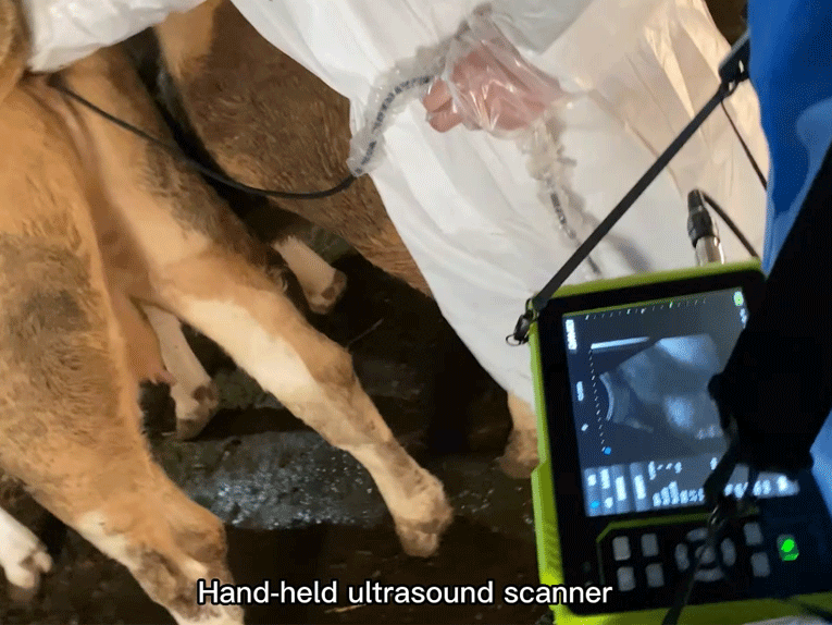
According to livestock reports in Indonesia, cows in tropical climates often experience ovarian inactivity due to heat stress. By combining ultrasound monitoring with hormonal protocols, breeding rates within 60 days postpartum have risen to 75%. Veterinarians using portable, waterproof, and user-friendly devices like the Dawei Y6 portable vet ultrasound machine have successfully implemented postpartum uterine health assessments in under-resourced areas, greatly improving the reproductive efficiency of small-scale farmers.
Veterinary ultrasound has become a global standard for managing postpartum recovery in cattle. By integrating advanced technologies like the Dawei Y6 handheld bovine and equine ultrasound machine, farmers and veterinarians can ensure more accurate assessments, faster diagnoses, and ultimately, better fertility outcomes. Backed by international research from Australia, USA, UK, Indonesia, and Italy, ultrasound-assisted reproductive monitoring is revolutionizing cattle health management worldwide.
Post time: Jun-24-2025