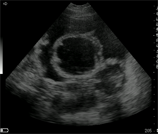
A 7.5 MHz probe is ideal for evaluating the ovaries and uterus in normal dogs, while a 10.0 MHz probe is used in cats. 5.0 MHz probes are useful for the diagnosis of mid- and late-stage pregnancies, uterine pus, and the detection of ovarian tumors. Padding blocks are useful for the exploration of small affected animals. The animal is often placed in a supine position, but can also be placed in a left or right lateral or standing position. Multi-position holding and multi-scanning surface exploration allows visualization of the entire reproductive tract. The best images are obtained by dehairing the lower abdomen. However, some owners object to pet hair clipping and have to scrub with ethanol or other aqueous agents to minimize the air between the probe and the skin before applying the coupling agent, thus improving image quality. Sonograms that are negative in early pregnancy should be repeated for several weeks of subsequent scans to confirm a false-negative diagnosis. Clipping is usually not required for diagnosis in mid- and late-term pregnancy, and may be withheld in cases of uterine pus and significant enlargement of the reproductive tract.
Ovarian localization is performed with longitudinal and transverse scans in the caudal and adjacent areas of the kidney. The ovaries may be in contact with the renal tail, or 2 cm posteriorly above it, posterolateral, posteromedial, or inferior to the renal tail. The sonographer is usually able to identify the ovaries on each side; in dogs and cats it may occur that the ovaries do not show up, due to their small size and the fact that they are often obscured by encapsulation of adipose tissue and overinflated intestines.
In addition, the ovaries have been removed in many animals. For uterine scanning, the bladder can be filled by drinking water, catheter introduction or drops of sterile water or saline to enhance uterine visualization. On the contrary, the bladder should be emptied before intravaginal scanning.7.2 Genital exploration techniques in males with 7.5 MHz or 10.0 MHz probe, 5.0 MHz or less can not provide sufficient resolution of the image, difficult to detect small lesions or microscopic parenchymal changes, it is generally believed that the frequency used for the appropriate resolution is very important to image the tissue structure to be in the focusing area of the probe. Transabdominal wall examinations are often used in small animal ultrasound imaging.
Image quality can be improved if an advanced endorectal probe is used, as it is not covered by other organs and has suitable short focusing properties. Intrarectal scanning is now used as a standard for prostate evaluation in humans and has been reported in dogs. Intrarectal scanners specifically for small animals will certainly appear in the future. As a rule, the lower abdominal coat is removed and a coupling agent is applied. However, in many animals the hair on the posterior lower abdomen is sparse and not removing the hair is sufficient to obtain good images, e.g., of the scrotum. Examination of the testes is generally done without clipping the hair, as there is a potential for irritation from clipping the hair, which can lead to injury to itself. Image quality can be improved both by using a padding block to keep small animal organs away from near-focal field artifacts, and by utilizing probe focusing.
In order to transect the abdomen from below, the supine position is usually used and scans are taken in various orientations to examine as many of the particular structures required as possible. Prostate localization is simple by placing the probe on the penile or prepuce side of the penis in the posterior lower abdomen, anterior to the pubic bone. Identification through the bladder is followed by a posterior scan, and once confirmed, it is carefully scanned in both longitudinal and transverse views. If located within the pelvic inlet, a frontal plane scan of the prostate may be required. A full bladder makes the prostate easier to visualize. A small amount of diuretic given during the examination will fill the bladder. Sterilized saline can also be instilled through a urinary catheter, but this method may produce nonspecific echoes by forming tiny air bubbles in the bladder. Tilting the examination table so that the tail of the affected animal is higher than the head can also move the prostate forward; it can also be done so that the tail of the affected animal is lower than the head, which facilitates filling and dilatation of the bladder triangle and urethral prostate visualization. Both areas must be scanned when examining the prostate. Testicular examination is relatively simple. The examination should be scanned in the transverse, longitudinal and frontal planes, and padding blocks often improve the quality of the image, allowing a better display of near-field structures.
Post time: Dec-19-2023


