


Principles of Small Animal Ultrasound Imaging
Small animal ultrasound probe will be ultrasound through the small animal skin surface coated coupling agent, ultrasound will be transmitted to the small animal body. Ultrasound waves undergo interfacial reflection when encountering the interface of two media with different densities, and the reflected back ultrasound waves are echoes, which are received by the ultrasound probe and formed into an ultrasound image after digital-mode conversion (Figure 1).
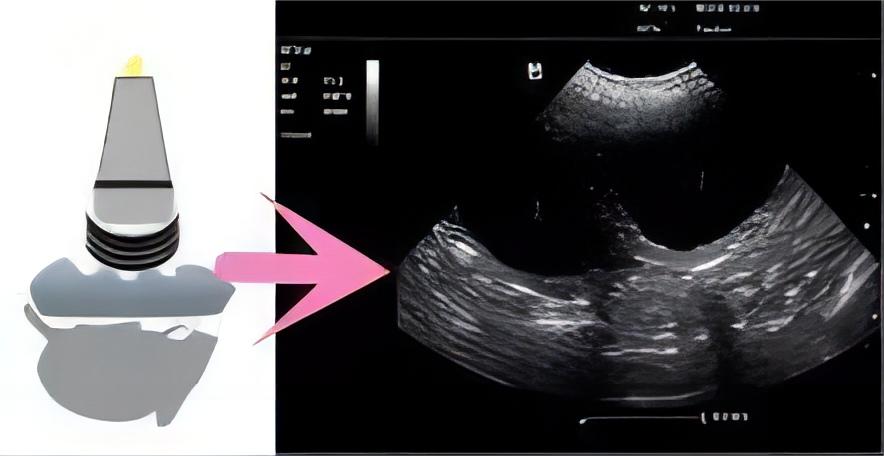
Figure 1
The basic imaging mode of ultrasound imaging is B-mode, this mode images the anatomical structure of small animals, displayed in a black-white-gray color scale, where:
White: represents strong echoes, generally dense tissue structures such as stones and air bubbles.
Gray: represents low echo, generally medium density tissue structures, such as liver, gallbladder, pancreas, spleen and other organs.
Black: represents no echo, generally low density tissue structures, such as fluid, blood vessels, necrotic tissue.
Another commonly used mode of ultrasound imaging is Color Doppler-mode, commonly known as color ultrasound, which is based on the B-mode structural image, the blood flow signals are marked with different colors, which is convenient to observe the distribution of blood flow in the tissues and organs, where:
Red: represents blood flow toward the probe (Figure 2, left).
Blue: represents blood flow back away from the probe (Fig. 2 right).
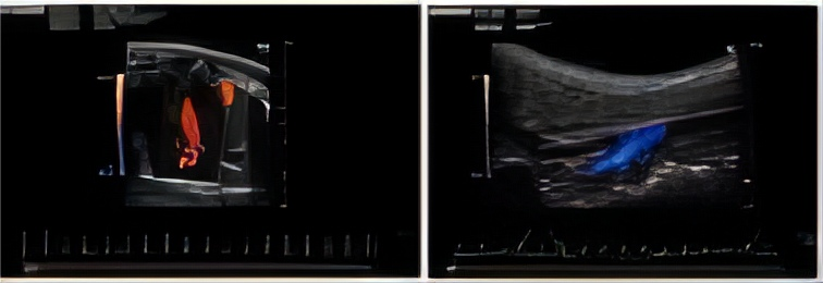
Figure 2
Small animal ultrasound imaging system features
1. Radiation-free, easy to operate, intuitive images, capable of real-time and long time duration observation (Fig. 3).
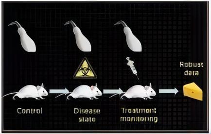
Fig. 3 Safe, non-invasive, long time duration study
2. Best at soft tissue imaging.
3. Wide field of application: in addition to the lungs imaging temporary difficulties (lungs filled with gas, in the ultrasound display as a strong echo region, can not see the internal structure), other tissues and organs can have ultrasound images.
Difference between clinical ultrasound and small animal ultrasound
The biggest difference between the two is the ultrasound probe frequency: clinical ultrasound probe frequency of about 3-15 MHz; small animal ultrasound probe frequency can generally reach 20-50 MHz, mouse ultrasound probe can reach 80 MHz.
According to the physical properties of ultrasound, the lower the ultrasound frequency, the better the depth of penetration, but the resolution becomes worse. Conversely, the higher the ultrasound frequency, the shallower the imaging depth, but the resolution increases. Therefore, most of the clinical ultrasound used is low-frequency ultrasound, which is suitable for the human body, and the image resolution obtained is sufficient for the observation of structures.
However, this is not the case for small animals. For example, the thickness of mice is only about 3 cm, and the volume of internal organs is much smaller than that of humans, so the use of ultrahigh-frequency probes to obtain high-resolution images in order to observe clearly (Figure 4).
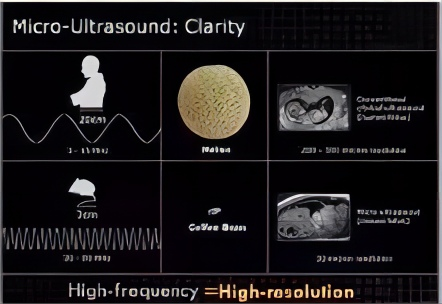
Figure 4 Difference between clinical ultrasound and small animal ultrasound imaging
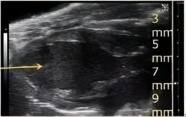
Take the mouse left ventricle ultrasound imaging as an example
The clinical ultrasound probe in Figure 5 has a low frequency and a deep imaging depth (about 3 cm), so the left ventricle of the mouse (less than 1 cm deep) cannot be placed in the center of the field of view, and the image resolution is not sufficient to
the left ventricle meticulously and analyze it accurately. The small animal ultrasound probe in Figure 6 has a high frequency, the left ventricle is located in the center of the field of view, and can be focused at a depth of 7mm, with good image resolution, which can clearly observe all important structures of the left ventricle, and is conducive to doing accurate quantification at a later stage.
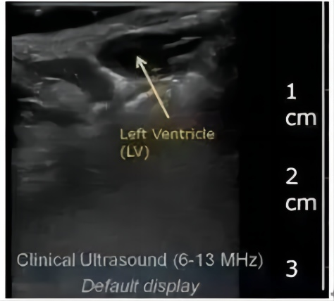
Figure 5 Clinical ultrasound imaging of the left ventricle in mice
Figure 6 Mouse left ventricle imaged by small animal ultrasound
Post time: Mar-13-2024