


The use of a porcine ultrasound machine to detect follicular dynamics and timing of ovulation in sows as part of herd reproduction management can constitute a valuable strategy to improve reproductive efficiency, as it allows to determine the optimal time to use for insemination, to greatly increase the rate of farrowing and litter size, and also to identify non-cyclic females (estrus).
Porcine ultrasound can be used for ovarian scanning by both the transrectal and transabdominal routes. Transabdominal scanning is performed by placing the transducer inferiorly below the mammary gland in the inguinal region in a manner similar to pregnancy diagnosis. The ovaries are located dorso-cranially relative to the posterior legs; the probe should be pointed upward toward the spine and tilted slightly anteriorly and posteriorly to visualize the bladder as a reference for locating the ovaries; in the ultrasound image, the ovaries will appear at the head of the bladder. When performed transrectally, the transducer is hand-held and manually guided through the rectum. The ovaries are located ventrally relative to the rectum and are approximately 30 cm to 40 cm internally. It is important to note that transrectal exams require prior fecal clearance to allow for sufficient tissue/probe interface to obtain high-quality imaging.
The transrectal approach is usually preferred because transabdominal scanning requires more practice to obtain good images.
The main limitation of transabdominal scanning with an ultrasound machine is related to the occlusion of the ovary by the surrounding tissue (i.e., the colon), especially on the left abdominal wall. In addition, it is often difficult to obtain detailed images of the ovarian structures due to the constant movement of the sow. On the other hand, transrectal scanning allows better and more detailed scanning of the ovary. It is even possible to calculate the ovarian structure.
In both techniques, fluid-filled follicles are shown as non-echoic ovarian structures of 3 mm – 11 mm during late estrus and estrus. Care should be taken to distinguish follicles from peripheral vessels, cysts, and hemorrhagic bodies. The echogenicity of the corpus luteum is similar to that of the ovarian stroma and can only be seen by trained researchers in about 50% of sows.
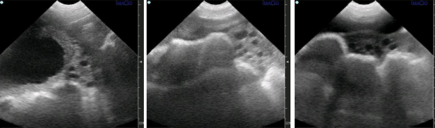
Ultrasound images of follicles prior to ovulation in the sow ovary for swine ultrasound abdominal testing
Repeated swine ultrasound examinations are required to determine the time of ovulation, and ovulation is thought to occur midway between the two investigative intervals in which a follicle was last detected and subsequently disappeared.
Post time: Feb-26-2024