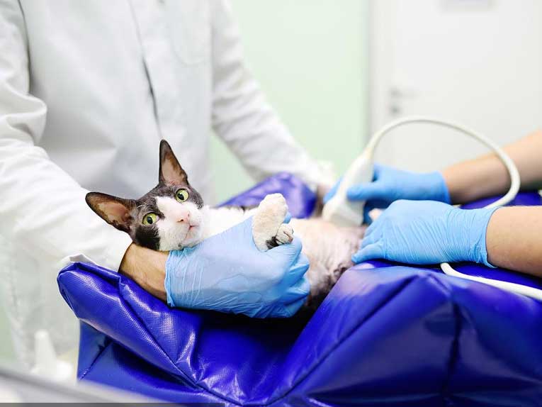
In the clinic, veterinarians often encounter urinary closure, urinary leaking, blood in the urine, polyuria and other clinical symptoms of dogs and cats, when encountering this situation, perhaps part of the clinical veterinarians in addition to laboratory tests such as urinary sediment, the first thought of diagnostic imaging may be the examination of animal X-rays, which may be related to the veterinarians on the degree of knowledge of veterinary X-rays and veterinary ultrasound diagnostic techniques or mastery of the degree of familiarity, for some clinical veterinarians For some clinical veterinarians, canine and feline ultrasound may be too abstract compared to animal X-ray examination, but from the clinical application of experience, in the examination and solution of this kind of problem, animal ultrasound diagnosis has an incomparable advantage of animal X-ray, which is because of their imaging principle determines their advantages. For example, in the orthopedic examination and chest examination, animal X-ray can easily present the corresponding lesion morphology, but in the bladder, the internal structure of the organs and the observation of pathological changes in the observation of the animal X-ray is indeed unable to help. However, the clinical diagnosis of animal ultrasound also has certain limitations, the presentation principle determines that the phenomenon of “a picture of many diseases, many diseases, one picture” is inevitable, so in the clinical application of veterinarians should be scanned in the image of the information combined with the results of other means of examination for a comprehensive analysis, identification and characterization.
I: Pet bladder ultrasound scanning section
Pet animal ultrasound sonogram is a certain thickness of information superimposed on the processing of a cross-section, coupled with some of the physical characteristics of ultrasound, according to the principle of ultrasound imaging, in the clinical application of some artifacts may inevitably appear (such as: volume effect, reverberation effect, rear enhancement effect, etc.), so this requires us to do any organ ultrasound to do multi-angle, multi-section of the target organ to observe. Therefore, this requires us to observe the target organ from multiple angles and multiple views when doing ultrasound examination of any organ, so ultrasound examination of the bladder is the same, at least two views (transverse plane, sagittal plane) glide scanning with fan scanning multi-angle multi-directional observation (this requires that the number of views should be far more than the number of views required by some references to the examination of the bladder), and as far as possible, through the observation of the two to three times after the spatial visualization of the brain to see a three-dimensional structure of the organ. The bladder should be examined in as many planes as possible, and as many planes as possible.
II: Pet bladder ultrasound observations
Some sources require that at least six views of the bladder be observed, including the bladder apex, the bladder body, and the bladder neck, because there is a preference for bladder lesions in these locations (e.g., cystitis in the bladder apex, tumors in the bladder neck, polyps in the bladder body, etc.), but of course, these lesion locations are not absolute. In any case, however, the observation of the sonogram during a cystoscopy is nothing more than an evaluation and analysis of the integrity and smoothness of the bladder wall, the thickness of the bladder wall and the contents of the bladder. The assessment of bladder wall integrity (whether the bladder wall is ruptured or not, it is important to note that a ruptured bladder wall usually has no or small amount of urine in the bladder, of course, I have also encountered a case of rupture of the bladder, but the bladder is still moderately full) and smoothness (mainly due to the inflammatory changes in the bladder wall, polyps, tumors and other pathologies that can lead to the unevenness of the wall of the bladder) is relatively simple, and is here to emphasize it. The assessment of thickness and bladder content is briefly described here.
First of all, the evaluation of the thickness of the bladder wall must have a holistic view, and the thickness of the bladder wall should be observed as a whole (the best state for cystometry is the semi-filled state of the bladder, and diuretics can be considered for oliguria, but this method is ineffective in cases of severe cystitis), because the bladder is a cystic organ, and the thickness of the bladder wall changes with the state of filling, so measuring the thickness of the bladder wall is the best way to determine whether or not the bladder is changing. Therefore, measuring the thickness of the bladder wall to determine whether the bladder has changed or not has some limitations (unless the thickening of the bladder wall is very obvious), therefore, we can measure the thickness of the bladder wall on the basis of the overall observation, which may be more able to increase the scientific validity of the results and persuasive.
Secondly, the changes in the bladder contents, the changes in the bladder can be divided into two cases of attachment to the bladder wall and the presence of the bladder lumen; abnormal structures attached to the bladder wall are usually common in bladder polyps or bladder tumors (lymphomas, migratory epithelial cell carcinoma, etc.). In pet ultrasound sonograms, bladder polyps usually show smooth and neat borders, whereas tumor projections usually show polymorphic borders and invasiveness to the bladder wall structures, and bladder tumors are usually accompanied by hematuria in clinical symptoms, whereas polyps are usually detected during physical examination and do not usually show abnormal clinical symptoms. elastography for tumor evaluation and improving tumor
Post time: Jan-04-2024


