


Introduction: Mobile C-Arms Bring Real-Time Imaging to Veterinary Surgery
The landscape of veterinary surgery is undergoing a profound transformation, shifting from an era defined by anatomical estimation to one of precise, visualization-driven intervention. For decades, veterinary surgeons have relied heavily on static pre-operative radiographs and tactile feedback to navigate complex procedures. Whether performing intricate fracture repairs or extracting foreign bodies, the traditional approach often forced surgeons to operate with limited visual confirmation, necessitating frequent interruptions to capture portable X-rays. This legacy model not only prolongs anesthesia duration—thereby increasing patient risk—but also struggles to meet the rising demand for minimally invasive techniques. Today, the integration of the veterinary mobile C-arm into modern animal hospitals is revolutionizing this paradigm, bringing technology once reserved for human medicine directly to the veterinary operating theater.
The adoption of these advanced systems marks the official entry of veterinary care into the age of “real-time imaging.” Unlike standard radiography which provides only a static snapshot in time, the latest C-arm systems offer real-time fluoroscopy for pets, effectively granting surgeons “X-ray vision” during active procedures. This capability allows clinical teams to visualize the exact trajectory of a guide wire, the reduction of a fracture gap, or the deployment of a stent as it happens. The immediate visual feedback loop eliminates the guesswork associated with blind manipulation, significantly enhancing surgical precision while reducing operative trauma. From spinal surgery to interventional cardiology, this continuous imaging capability has evolved from a luxury add-on to a critical necessity for high-standard care.
Furthermore, the quality of intraoperative imaging has achieved a new benchmark. Modern devices, such as the RCM-605XV, utilize cutting-edge dynamic flat-panel detectors to deliver high-resolution veterinary imaging that rivals diagnostic radiology suites. These systems are capable of resolving minute trabecular bone structures and tiny vascular branches with exceptional clarity, even while the anatomy is in motion. By overcoming the distortion and blooming artifacts common in older image intensifier technology, these next-generation C-arms empower veterinarians to make confident, evidence-based decisions instantly. This shift from merely “seeing” to “clearly distinguishing” fine details is fundamentally improving safety outcomes and success rates in complex surgical cases.
Why Mobile C-Arms Are Becoming the Clinic Standard
In the high-stakes environment of a modern veterinary operating room, the limitations of traditional Digital Radiography (DR) are becoming increasingly apparent, particularly for complex orthopedic and interventional cases. Standard DR is inherently a static “snapshot” technology; to verify the placement of an implant or the alignment of a bone, the surgeon must physically halt the procedure, reposition the equipment (or the patient), capture an image, and wait for processing. This stop-and-start workflow disrupts surgical rhythm and cumulatively extends anesthesia time, which is a critical risk factor for animal patients. Moreover, static images fail to capture dynamic physiological processes or the real-time interaction between instruments and tissue, leaving a blind spot during critical maneuvers like catheter navigation or joint articulation.
veterinary mobile C-arm
By contrast, a mobile C-arm provides continuous fluoroscopic guidance, capturing dozens of frames per second to create a live video feed of the internal anatomy. This real-time fluoroscopy for pets is indispensable for procedures requiring high precision. In intramedullary nailing, for instance, a surgeon can watch the guide pin advance through the marrow cavity in real-time, adjusting the angle instantly to avoid breaching the cortex. Similarly, during tracheal or esophageal foreign body retrieval, live imaging reveals the object’s relationship to surrounding soft tissues, allowing for safe extraction without damaging vital structures. Industry analysis suggests that clinics equipped with intraoperative fluoroscopy can reduce surgical time for complex fractures by over 30%, significantly lowering infection risks and improving recovery prognoses.
The following table outlines the critical operational differences between relying on traditional static DR versus utilizing a modern mobile C-arm during surgery:
| Feature | Traditional Intraoperative DR | Mobile C-Arm (Fluoroscopy) |
| Temporal Feedback | Delayed (Pause, Shoot, Wait) | Instant (Real-time, Continuous) |
| Dynamic Motion | None (Static Capture) | Full Motion (Contrast flow, Joint movement) |
| Multi-Angle View | Difficult (Must move patient) | Flexible (Rotate C-arm around patient) |
| Primary Use Case | Diagnosis, Post-op Verification | Intraoperative Navigation, Interventional |
RCM-605XV Key Technical Specifications and Clinical Implications
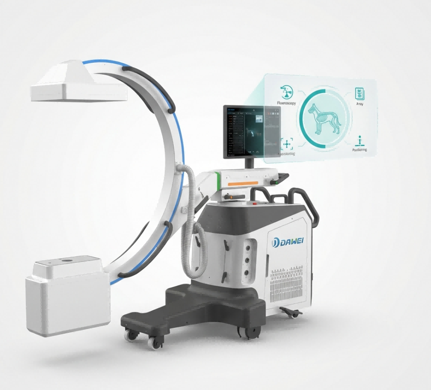
The RCM-605XV system represents a significant leap forward in veterinary imaging technology, engineered specifically to address the unique anatomical and procedural challenges of animal healthcare. At the heart of its imaging chain lies a state-of-the-art Amorphous Silicon Dynamic Flat Panel Detector. Unlike older image intensifiers that suffer from geometric distortion at the edges, this flat panel technology ensures uniform, distortion-free imaging across the entire field of view. According to the RCM-605XV technical specifications, the detector features a large active area of 204mm × 204mm (approx. 21cm × 21cm), which is sufficient to cover the thoracic cavity of a medium dog or bilateral joints in a single view, reducing the need for repositioning.
Precision in imaging is further guaranteed by the system’s high-density pixel matrix. The detector boasts a 1024 × 1024 matrix with a pixel pitch of just 200 μm. This granular level of detail is critical for identifying hairline fractures or verifying the precise deployment of micro-stents. Supporting this high-resolution capture is a powerful 5kW High-Frequency X-ray Generator, capable of a broad exposure range from 40kV to 125kV. This wide dynamic range allows veterinarians to image patients varying vastly in size—from a 500g kitten to a 60kg Mastiff—with equal clarity. The system’s “Pulse Fluoroscopy” mode is a key safety feature, delivering high-quality images while significantly reducing the cumulative radiation dose to both the patient and the surgical team.
RCM-605XV Technical Specifications Overview
|
High Resolution Meets Real-Time: Efficiency and Safety
|
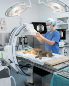
The vertical form factor of the mobile C-arm allows for flexible positioning in tight surgical spaces. |
In veterinary surgery, speed and clarity are not just matters of convenience—they are pillars of patient safety. The RCM-605XV system delivers on both fronts by combining high-speed acquisition with exceptional image depth. The system operates at a high frame rate of 30 fps (frames per second), which creates a seamless, movie-like visualization of internal anatomy. This high temporal resolution eliminates the “ghosting” or motion blur often seen in lower-end systems. When a surgeon is manipulating a catheter within a beating heart or reducing a fracture in a trembling muscle, the display updates instantly, ensuring that hand movements are perfectly synchronized with the visual feedback on the screen.
Equally important is the system’s ability to render subtle tissue differences. With a 16-bit AD conversion capability, the detector can differentiate over 65,000 shades of gray, far surpassing the 12-bit or 14-bit depth of standard equipment. This high dynamic range means that dense bone and soft tissue can be visualized clearly in the same image without overexposure or underexposure. Combined with a spatial resolution of 2.5 lp/mm, surgeons can confidently identify the finest details, such as the tip of a 0.014-inch guide wire. Furthermore, the intelligent Auto Brightness Control (ABC) automatically adjusts radiation parameters in milliseconds as the C-arm moves over different body thicknesses, guaranteeing consistent image quality so the surgical team can focus entirely on the patient.
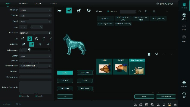
The intuitive workstation interface allows for rapid patient setup and anatomical protocol selection. |
Clinic Implementation Guide: From Selection to Daily Use
Introducing a veterinary mobile C-arm into a general practice or specialty clinic involves more than just plugging in a machine; it requires a thoughtful approach to workflow and safety. To maximize the return on investment and clinical efficacy, practices should consider the following implementation steps:
- Surgical Suite Layout & Geometry: While modern C-arms are compact, they require specific spatial clearances to function effectively. The RCM-605XV features a generous opening of ≥870mm, which provides excellent clearance for bulky drapes and large patients. However, clinics must ensure a clear radius of at least 1.5 meters around the surgical table to allow the C-arm to rotate and slide freely. Level, anti-static flooring is also crucial for smooth, precise positioning of the unit.
- Team Choreography & Training: Efficient C-arm use is a team sport. It typically requires coordination between the sterile surgeon and a non-sterile assistant who manipulates the C-arm locks and controls. Clinics should run dry simulations of common procedures (e.g., “simulate a lateral view for a radial fracture”) to establish clear verbal commands and smooth mechanical movements. A well-trained team can position the C-arm in seconds, drastically reducing anesthesia time.
- Radiation Safety Protocol: Safety is paramount. The RCM-605XV is designed with low leakage (< 0.5 mGy/h), but standard ALARA (As Low As Reasonably Achievable) principles apply. All staff must wear lead aprons and thyroid shields. Crucially, operators should utilize the “Pulse Fluoroscopy” mode whenever possible; this pulses the X-ray beam rather than keeping it continuous, cutting total radiation dose significantly without compromising diagnostic quality.
- Procedural Integration: Start with high-value, high-frequency procedures. Use the C-arm to verify alignment in fractures before closing, or for minimally invasive cystoscopy. Leveraging the orbital rotation allows for checking orthogonal views (90-degree separation) without flipping the patient, which preserves the sterile field and fracture reduction.
Frequently Asked Questions (FAQ)
For clinics considering their first C-arm purchase, here are answers to the most common questions regarding clinical utility and ownership:
Q: We already have a DR system. Why do we need a mobile C-arm? A: DR answers “What is it?” (Diagnosis), while a C-arm answers “Where am I?” (Navigation). DR is a static verification tool, whereas a C-arm is a dynamic guidance tool. They are complementary; the C-arm allows you to perform surgeries that are impossible or high-risk with blind technique, such as closed reductions or vascular interventions.
Q: Is this system suitable for small exotic animals or kittens? A: Absolutely. The RCM-605XV has a wide energy range starting as low as 40kV. Combined with automatic exposure control, it can dial down the radiation output to capture high-contrast images of very small patients safely, without “burning out” the image.
Q: How do we manage radiation exposure during long surgeries? A: Through technology and technique. The system features “Last Image Hold,” which keeps the final fluoroscopic image on screen after the pedal is released, allowing the surgeon to study the anatomy without continuous exposure. Using pulse mode and collimation (narrowing the beam to just the region of interest) further reduces the dose.
Q: Can the images be saved to our practice management software? A: Yes. The workstation is fully DICOM 3.0 compliant. Both static spot films and dynamic video loops can be exported directly to your clinic’s PACS server or cloud storage for permanent archiving, client education, or specialist referrals.
Q: What is the maintenance schedule? A: Standard protocol involves a mechanical inspection and lubrication every six months to ensure smooth movement, and an annual physics inspection to calibrate image quality and verify radiation safety compliance.
Conclusion & Call to Action
The future of veterinary medicine is undeniably moving toward minimally invasive, precision-guided care. The veterinary mobile C-arm is the cornerstone of this evolution, empowering surgeons to see more, do more, and treat more effectively. By integrating real-time fluoroscopy for pets into your surgical workflow, you are not just upgrading equipment—you are elevating the standard of care, offering faster recoveries and better outcomes for your patients. With capabilities like high-resolution veterinary imaging, clinics can confidently expand their service offerings to include advanced orthopedics and interventional procedures.
If you are ready to transform your surgical capabilities, we invite you to experience the difference firsthand. Contact our specialist team today to schedule a demo of the RCM-605XV or to discuss how this technology can fit into your practice’s growth plan.
Post time: Feb-03-2026