


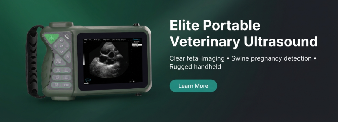
Introduction: What Is Swine Ultrasound Diagnosis?
Swine ultrasound diagnosis is a cornerstone of modern veterinary practice, offering a non-invasive window into the reproductive health of livestock. By utilizing high-frequency sound waves, this technology allows farm owners and veterinarians to visually confirm pregnancy, monitor fetal development, and detect uterine abnormalities with precision that traditional methods cannot match.
Implementing veterinary ultrasound for pigs on a farm leads to significant efficiency gains. Early detection of non-pregnant sows (“open” sows) reduces non-productive feed days, directly impacting the bottom line. Furthermore, pig pregnancy ultrasound provides immediate feedback, allowing for timely decision-making regarding herd management and health interventions. Whether for a smallholding or a large commercial operation, the ability to “see” inside the animal transforms reproductive management from a guessing game into a science.
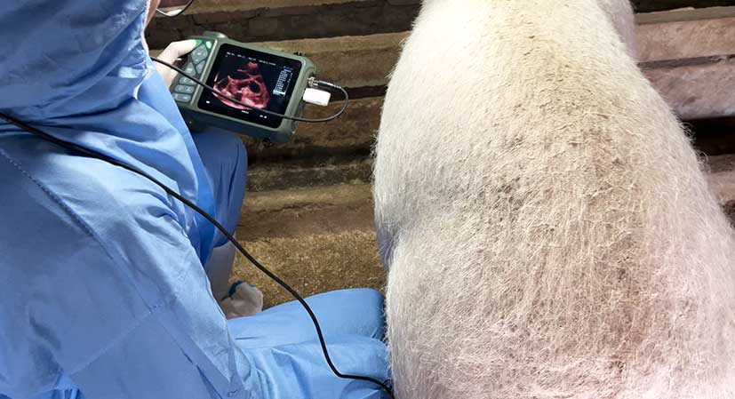
Veterinarian performing swine pregnancy ultrasound in a barn
Ultrasound Safety for Pigs
According to the Swine Ultrasound Guide, ultrasound technology is fundamentally safe for both the operator and the animal. Unlike X-rays, which use ionizing radiation, ultrasound relies on sound waves that have no known harmful effects on biological tissue when used for diagnostic purposes.
Safety Highlights
- Non-Invasive: The procedure requires no surgery or needles, minimizing stress for the sow.
- No Radiation: Completely free of radioactive substances, making it safe for repeated use.
- Fetal Safety: Proven safe for pregnant animals and unborn piglets; widely used in human medicine for the same reason.
Common Safety Questions
- Q: Is there any risk to the operator?
A: No, there are no known side effects or radiation risks for the person performing the scan.
- Q: Can it induce stress in the animal?
A: The process is painless. With proper handling, stress is negligible compared to invasive procedures.
Why Ultrasound Beats Traditional Methods
For decades, farmers relied on visual observation or external palpation to guess at pregnancy status. Ultrasound replaces these subjective methods with objective data. The visual clarity provided by swine pregnancy detection devices eliminates ambiguity.
Traditional Methods
- Palpation: Subjective and difficult; cannot confirm fetal viability.
- Blood Tests: Accurate but slow; requires lab time and incurs recurrent costs per test.
- Visual Wait: Costly due to wasted feed if the sow is not pregnant.
Ultrasound Benefits
- Visual Confirmation: Detects fetal heartbeat, skeletal structures, and movement.
- Diagnostic Depth: Can identify abscesses, pus, or signs of fetal demise.
- Cost Efficiency: After equipment purchase, the cost per scan is negligible.
How Pig Ultrasound Works: Basics and Image Quality
Understanding the physics of ultrasound ensures better results. The system works on the echo principle: the transducer (probe) emits high-frequency sound waves into the body. These waves travel through tissues and bounce back when they hit interfaces between different tissue densities. The device processes these returning echoes to form a black-and-white image on the screen.
Image Quality Factors:
- Liquid & Soft Tissue: Ultrasound penetrates these easily, appearing dark or black (e.g., fluid in the bladder or embryonic sacs).
- Bones & Gas: Ultrasound cannot penetrate bone or gas. Bones appear as bright white lines with shadows behind them, while intestinal gas can scatter the beam, causing a “cloudy” or poor-quality image.
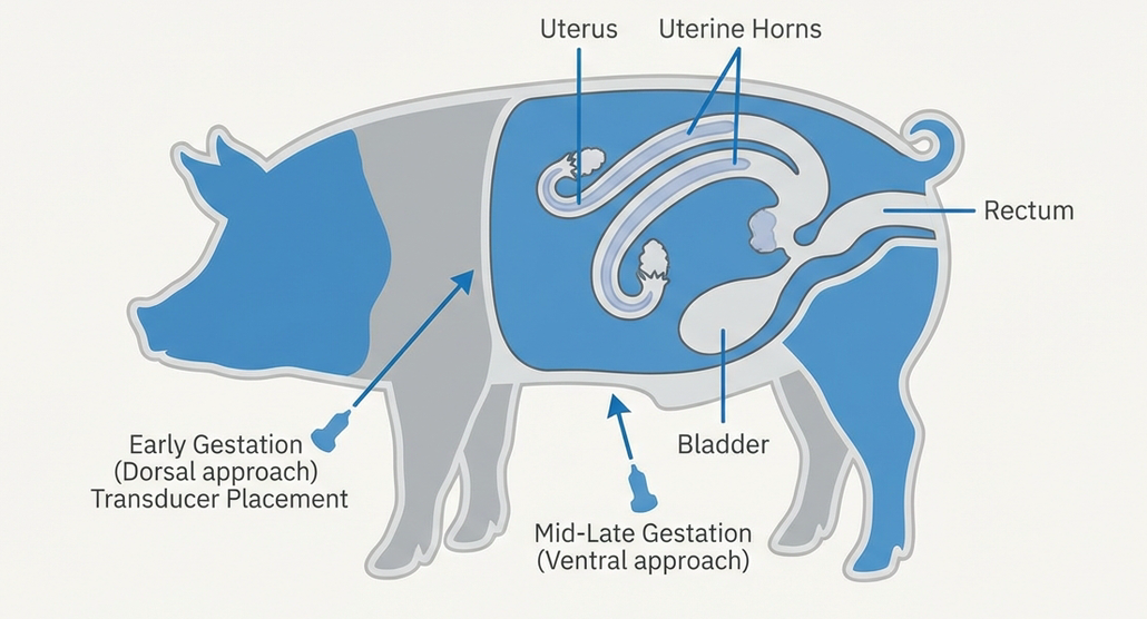
Recommended probe placement and uterus anatomy (illustration)
Step-by-Step: Performing a Pig Pregnancy Ultrasound
Performing a scan requires technique and knowledge of anatomy. Follow this protocol for accurate pig pregnancy ultrasound diagnosis.
Preparation
Ensure the animal is calm. Apply a generous amount of coupling gel to the probe. The gel is critical as it eliminates air gaps between the probe and skin, ensuring sound waves can enter the body.
Positioning
Ideally, scan while the sow is standing or feeding. Locate the flank area near the rear leg, avoiding the rib cage.
Probe Placement (Critical)
For early pregnancy (up to 60days), place the probe slightly above the second-to-last pair of udders. Angle the probe 45° toward the spine and forward toward the pig’s head. For later pregnancy (after 60 days), move to the third or fourth-to-last pair of udders to account for the uterus dropping.
Interpretation
Look for key indicators. A non-pregnant uterus appears as a bright line near the intestine. Pregnancy is confirmed by black fluid-filled sacs (25 days), calcified white bones (55 days), or distinct ribs and spine (90 days).
Troubleshooting and Best Practices
Even experienced technicians encounter unclear images. The Swine Ultrasound Guide suggests specific remedies for common issues. The most frequent culprit is intestinal gas, which blocks sound waves. If the image is cloudy, try repositioning the probe or scanning a different area.
Quick Troubleshooting Checklist
- Gel: Is there enough coupling gel? Dry skin blocks signals.
- Bone Interference: Are you hitting the pelvic bone or ribs? Adjust your angle to 45°.
- Landmarks: Can you find the bladder? This large liquid-filled area (dark on screen) is your best landmark to orient yourself.
- Settings: Is the gain/brightness adjusted correctly for the ambient light in the barn?
Clinical Use Cases and Examples
Real-world application involves checking for viability and health, not just presence.
1. Confirmation Windows:
The ideal window for confirmation is 25–30 days post-mating. At this stage, fetal sacs are clearly visible as black circles.
2. Fetal Viability:
Around 90 days, you can observe the fetal heart. A healthy fetal heart rate is approximately 182 beats/minute. Detecting this confirms the piglet is alive and thriving.
3. Uterine Abnormalities:
Ultrasound can also identify uterine inflammation (endometritis). Unlike the clean black of pregnancy fluid, inflammation appears as irregular “strips” of liquid or mass-like echoes within the uterine horn.
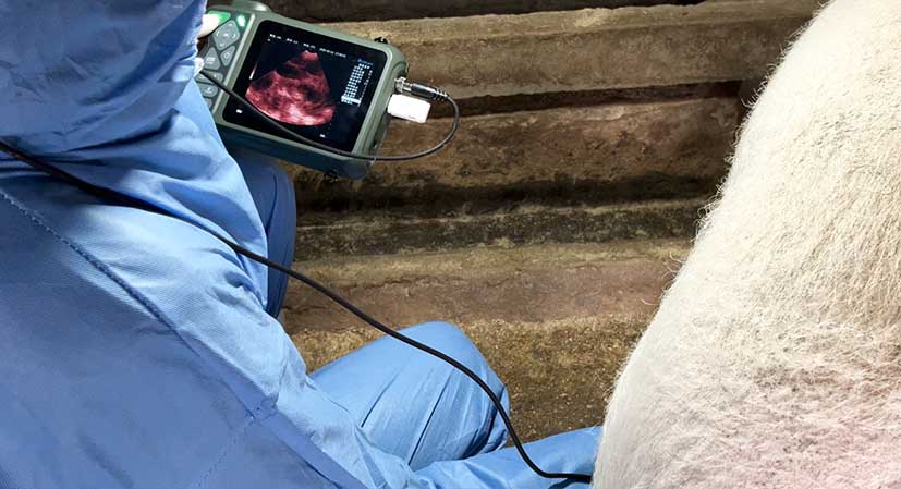
On-farm swine ultrasound in practice.
Ready to upgrade your farm’s reproductive management?
Contact your veterinary equipment supplier today for training and the latest in portable swine ultrasound technology.
Post time: Jan-27-2026