


What is veterinary color Doppler ultrasound?
Veterinary color Doppler ultrasound is a medical imaging technique used in veterinary medicine to assess blood flow within the body of an animal. It is an advanced form of ultrasound that provides real-time, dynamic images of blood vessels and blood flow patterns, allowing veterinarians to evaluate the circulatory system and diagnose various conditions. Unlike potentially dangerous x-rays, ultrasound is considered safe.
Ultrasound machine directs a narrow beam of high-frequency sound waves to the area of interest. The sound waves can pass through, reflect, or absorb the tissue they encounter.
The reflected ultrasound waves are returned to the probe as “echoes” and are converted into an image.
The reflected ultrasound waves are returned to the probe as “echoes” and converted into an image that is displayed on a monitor, giving a two-dimensional “picture” of the tissue being examined.
This technique is invaluable for the examination of internal organs and is being used for the first time in veterinary medicine for the diagnosis of pregnancy. However, this technique is also very useful in evaluation. Changes in cardiac conditions abdominal organs. Ultrasonography is useful in the diagnosis of cysts and tumors.
Are there any disadvantages to this technique?
“Ultrasound does not pass through air.”
Ultrasound has little value in examining organs that contain air. Ultrasound waves will not pass through air, so it cannot be used to examine normal lungs. Bones also block ultrasound, so the brain and spinal cord cannot be examined by ultrasound and, obviously, bones cannot be examined.
Are there different forms of ultrasound?
Ultrasound can take various forms depending on the image produced. In veterinary practice, the most common form of ultrasound (brightness mode) is two-dimensional ultrasound. This gives a two-dimensional image of the scanned organ. This is a type of ultrasound used for examining abdominal structures, making pregnancy diagnoses, evaluating heart function, and examining eye diseases.It is used to examine abdominal structures, diagnose pregnancy, assess cardiac function and examine the eyes.M-mode (motion mode) is a B-mode in which motion tracking of the structure being scanned is displayed. The walls, chambers, and valves of the heart are examined using a combination of M-mode and 2D ultrasound to evaluate cardiac function. Cardiac ultrasound is often referred to as echocardiography. Doppler ultrasound is a specialized form of cardiac ultrasound that measures the direction and speed of blood flow through the heart and blood vessels. Color Doppler technology has made it easier to observe the flow of blood through the heart and vital blood vessels.
Does my dog need anesthesia?
“Anesthesia is usually not needed; this technique is completely painless.”
Most ultrasounds usually do not require anesthesia unless a biopsy is to be performed. The technique is completely painless and most dogs will lie comfortably while the scan is being performed. Occasionally, if the dog is very fearful or cranky, sedation may be required.
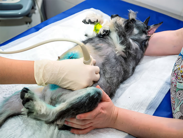
Is it necessary to shave my dog?
In most cases, the fur must be shaved in order for the ultrasound to be performed. Since ultrasound is not transmitted through the air, the handheld probe must be in complete contact with the skin. In some cases, such as pregnancy diagnosis, it may be possible to get adequate images by moisturizing the hair with alcohol and applying a generous amount of water-soluble ultrasound gel.
“Ultrasound images will have better quality if the area to be examined is shaved.”
However, in all cases, the ultrasound image will be of better quality if the area to be examined is shaved.
When will I know the results of the examination?
Since ultrasound studies are performed in real time, the results seen are known immediately. In some cases, ultrasound images can be sent to a veterinary radiologist for further consultation. When this happens, the final report may not be available for several days.
Is this technology affordable?
While the initial cost of the scan may seem high, it must be equated to the high cost of the equipment, the need for specialized training to interpret the images, and the amount of time it takes to perform the exam. It is useful for pregnancy diagnosis, evaluation of internal organs, evaluation of cardiac function, and evaluation of certain ocular diseases, making it an invaluable, non-invasive diagnostic tool to help protect your pet’s health.
Post time: Jan-10-2024