


A customer case showing how the RV-32A veterinary digital radiography system can support veterinary imaging education, practical training, and digital X-ray workflow optimization at Shenyang Institute of Technology.
Customer Case: Shenyang Institute of Technology
Product Used
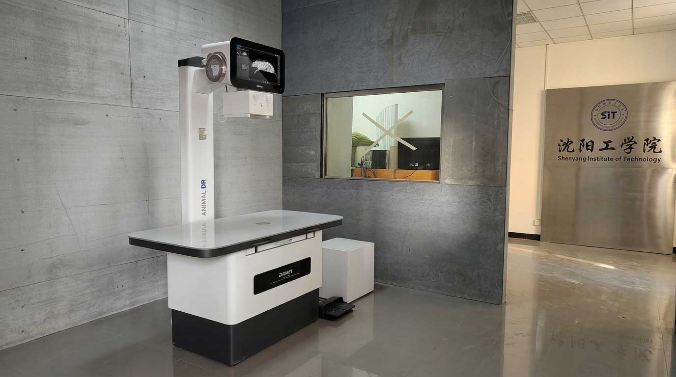
RV-32A Animal Digital Radiography System
Customer Type
University / teaching institution / laboratory and practical training environment
Application Scenario
Veterinary imaging education, practical radiography training, small-animal examination workflow, and digital X-ray image management
Executive Summary
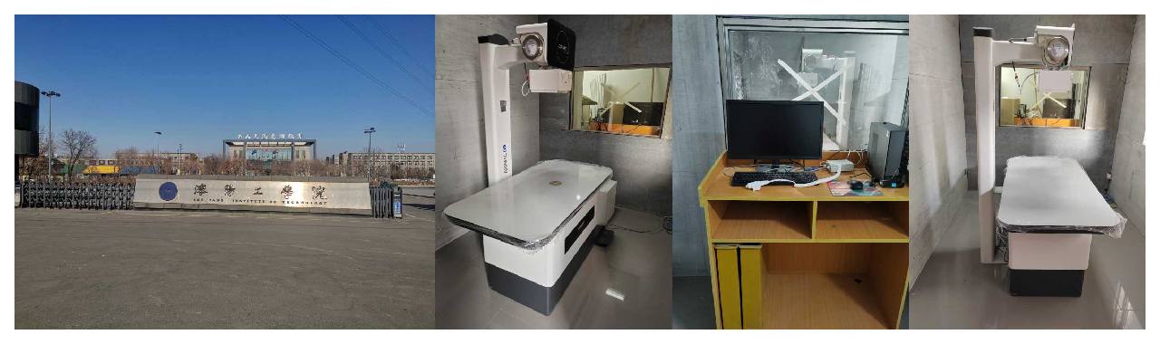
Shenyang Institute of Technology is known for its practice-oriented education model and laboratory-based teaching environment. Publicly available university information shows that the institution operates multiple experimental teaching centers and laboratories, making it a suitable customer profile for advanced teaching and imaging equipment deployment.
In this customer case, the RV-32A animal digital radiography system is positioned as a practical solution for institutions that need more than simple X-ray acquisition. In a university or training environment, the system can help combine image quality, equipment stability, workflow efficiency, and teaching usability in one platform. For schools and training centers that need to improve demonstration quality, student practice experience, and image review efficiency, the RV-32A offers a strong application fit.
Project Background
In teaching institutions and practical training environments, animal imaging equipment is expected to do more than generate diagnostic images. It must also support teaching demonstration, operator training, repeatable positioning practice, and efficient image review. For a customer such as Shenyang Institute of Technology, this means the equipment must perform well not only technically, but also operationally.
A conventional imaging workflow can become inefficient when image acquisition is slow, positioning space is limited, or the equipment interface is not intuitive enough for teaching use. In practical classes or skills training sessions, instructors often need to explain positioning logic, exposure workflow, and image interpretation while students observe or participate. If the system is slow to respond or difficult to operate, it can reduce teaching efficiency and interrupt the rhythm of practical instruction.
Against this background, the RV-32A offers a more modern animal DR workflow. Its integrated structure, large imaging area, flat panel detector, and fast image output characteristics make it well suited for animal radiography scenarios where both performance and training usability matter.
Project Challenges
1. Teaching Environments Need Stable and Repeatable Imaging Workflow
In a university or training setting, the imaging system is often used by multiple operators under instructor guidance. That means the workflow needs to be stable, easy to understand, and repeatable across different sessions. A complicated operation process can increase teaching difficulty and reduce hands-on efficiency.
2. Practical Training Requires Fast Feedback
In radiography training, immediate image feedback is extremely important. Instructors need students to see positioning results quickly so they can correct posture, exposure logic, and image interpretation in real time. If image output is delayed, the learning loop becomes slower and less effective.
3. Different Animal Imaging Tasks Require Adequate Coverage and Resolution
Animal radiography in teaching and routine examination may involve different body sizes and anatomical regions. The system therefore needs a sufficiently large effective imaging area, reliable detector performance, and clear image detail to support both demonstration and practical use.
4. Equipment Must Fit a Structured Teaching Space
Teaching institutions value orderly workflow and physical usability. Bed size, load-bearing capacity, detector integration, and table movement all affect how comfortably the system can be used in a lab or training room. If the system is not well designed for practical operation, it may limit positioning efficiency and reduce teaching fluidity.
Why the RV-32A Was a Good Fit
The RV-32A is designed as an animal digital radiography solution that combines a high-voltage generator, X-ray source assembly, flat panel detector, collimator, and mechanical table structure into a practical clinical and teaching system.
Based on the provided technical data, the system brings together several strengths that are highly relevant to an education and training customer:
- 32 kW high-voltage generator for stable imaging performance
- Tube voltage range of 40-150 kV and tube current range of 10-400 mA
- Amorphous silicon flat panel detector with CsI scintillator
- 16-bit A/D conversion for digital image acquisition
- 430 mm × 430 mm effective imaging area
- 3072 × 3072 acquisition matrix with 140 μm pixel size
- Image output time of ≤1 second for faster feedback
- Spatial resolution of 3.6 LP/mm
- LED collimator for positioning assistance
- Four-way floating flat table with electromagnetic lock
- Table size of 1400 mm × 720 mm and uniform load capacity of ≥100 kg
These features make the RV-32A especially attractive for institutions that need both teaching practicality and real imaging capability.
How the RV-32A Can Support the Customer
1. Improve Veterinary Imaging Demonstration in Teaching
The RV-32A can help instructors present digital animal radiography more clearly in classroom and practical teaching scenarios. With a structured table design, integrated imaging workflow, and rapid output, it is easier to demonstrate positioning logic, exposure steps, and image acquisition results in a way that students can follow.
2. Support Faster Hands-On Training Cycles
Because the detector can deliver images in ≤1 second, the system supports faster practice feedback. Students and instructors can review the result soon after exposure, making it easier to adjust positioning and improve operational understanding during the same session.
3. Provide More Practical Coverage for Animal Imaging Tasks
The 430 mm × 430 mm imaging area and 3072 × 3072 matrix help the system handle a broad range of animal radiography tasks. In a training environment, this is valuable because one platform can support multiple teaching examples rather than being limited to a narrow use case.
4. Make the Workflow Easier to Standardize
The physical design of the RV-32A also contributes to usability. The four-way floating flat table, adequate load capacity, and structured system layout can help instructors and operators build a more standardized examination process. In a teaching context, standardization is important because it improves consistency from one training session to the next.
5. Support Digital Image Review and Teaching Discussion
A digital DR workflow does not stop at image acquisition. It also creates opportunities for image comparison, post-exposure review, case discussion, and teaching explanation. In a university context, this makes the RV-32A valuable not only as an imaging device, but also as a teaching support tool.
Implementation-Oriented Value
For a customer like Shenyang Institute of Technology, the value of the RV-32A can be understood through the actual teaching and practical workflow it supports.
First, the system can function as a training-oriented digital radiography platform for animal imaging education. It helps bridge the gap between theory and hands-on practice by giving students more immediate visual feedback and a more structured operating process.
Second, the RV-32A can support a more efficient practical class rhythm. In teaching environments, repeated waiting or frequent repositioning confusion can waste valuable instruction time. A system with fast image generation and clear physical workflow is better aligned with training efficiency.
Third, the system supports a more professional learning experience. Instead of training on outdated or limited imaging setups, students can engage with a modern DR-based workflow that is closer to current clinical and institutional imaging practice.
Project Results
More Efficient Practical Training Workflow
By enabling rapid digital image output and a more structured operating sequence, the RV-32A can help teaching sessions move more smoothly. Instructors can spend less time waiting for image feedback and more time explaining positioning, anatomy, and image interpretation.
Better Student Understanding Through Immediate Visual Feedback
Fast image generation makes it easier for students to connect positioning actions with image results. This strengthens the teaching loop and supports more intuitive learning during radiography practice.
Stronger Fit for Institutional Imaging Environments
With its integrated generator, detector, collimator, and table structure, the RV-32A provides a more complete imaging platform for institutions that need both practical usability and dependable technical performance.
Greater Value Beyond a Single Imaging Task
For a university customer, the benefit of the RV-32A is broader than one-time equipment installation. It can support demonstration teaching, routine practice, skills training, image review, and academic discussion, making it a more versatile long-term asset.
Technical Highlights of the RV-32A
- 32 kW output power
- 40-150 kV tube voltage range
- 10-400 mA tube current range
- 1.0 mm / 2.0 mm focal spot
- 900 kJ tube heat capacity
- Amorphous silicon detector with CsI scintillator
- 16-bit digital acquisition
- 430 mm × 430 mm effective imaging area
- 3072 × 3072 image matrix
- 140 μm pixel size
- ≤1 second image output
- 3.6 LP/mm spatial resolution
- LED collimator
- Four-way floating flat table
- ≥100 kg table load capacity
FAQ
What is the RV-32A animal digital radiography system used for?
The RV-32A is used for digital X-ray imaging in animal radiography scenarios. It can support veterinary examination, imaging training, teaching demonstration, and practical DR workflow in educational or clinical environments.
Why is the RV-32A suitable for veterinary teaching institutions?
The system combines fast image output, a large detector area, stable DR performance, and a practical table structure. These features make it useful for instructor-led demonstration, student training, and repeated imaging practice.
How fast is the RV-32A image output?
According to the provided specifications, the detector image output time is ≤1 second, which is valuable for practical training and workflow efficiency.
What detector size does the RV-32A use?
The system uses a flat panel detector with an effective imaging area of 430 mm × 430 mm, supporting a broad range of animal imaging tasks.
What are the key advantages of the RV-32A for digital animal radiography?
Its main advantages include 32 kW output power, digital flat panel imaging, fast image generation, structured table operation, and suitability for both imaging performance and teaching usability.
Conclusion
This case shows how the RV-32A animal digital radiography system can be positioned for a university customer that values practical teaching, laboratory usability, and modern imaging workflow. For institutions such as Shenyang Institute of Technology, the system offers a strong combination of DR performance, operational clarity, and training value.
Whether the goal is to improve veterinary imaging instruction, strengthen practical radiography education, or modernize animal X-ray workflow in a teaching environment, the RV-32A provides a capable and application-oriented solution.
Post time: Jun-26-2026