


In the rapidly evolving landscape of veterinary medicine, the demand for diagnostic precision and mobility has never been higher. For canine practitioners, the ability to perform advanced imaging at the point of care—whether in a busy clinic, a client’s home, or a remote rescue shelter—is a transformative capability. This brings us to the critical category of portable dog ultrasound machines under 5kg. These lightweight, powerful devices bridge the gap between heavy cart-based systems and basic handheld scanners, offering the perfect balance of diagnostic power and physical portability. Leading manufacturers like Dawei Veterianry Medical have recognized this need, developing sophisticated solutions that pack high-end features like Color Doppler and Tissue Harmonic Imaging into notebook-sized frames.
Why Lightweight Ultrasound Matters for Canine Practice
The shift towards “ultra-portable” veterinary equipment is not merely a trend but a response to the practical realities of modern animal healthcare. A device under 5kg changes the workflow significantly compared to traditional 20kg+ cart systems.
1. Mobile Veterinary Services
For mobile vets, every kilogram counts. Carrying equipment from a vehicle to a patient’s living room requires gear that is ergonomic and manageable. A laptop-style ultrasound allows practitioners to bring hospital-grade diagnostics directly to nervous or immobile dogs, reducing stress for the animal and owner.
2. Space-Constrained Clinics
Small urban clinics often lack the floor space for permanent imaging suites. A portable unit can be stored in a cupboard or carried between exam rooms, maximizing utility without occupying valuable square footage. The Color Doppler systems available today ensure that size does not compromise diagnostic confidence.
3. Emergency and Rescue Work
In emergency scenarios or rescue shelters, power availability and environmental conditions can be unpredictable. Lightweight units with integrated batteries and robust build quality allow for immediate FAST (Focused Assessment with Sonography for Trauma) scans to detect internal bleeding or cardiac issues in rescued dogs on-site.
When evaluating these systems, it is essential to look beyond just the weight. True portability involves a synergy of battery life, boot-up speed, and ruggedness. A sub-5kg machine that requires a constant mains connection or has a fragile chassis fails the portability test. Furthermore, the ergonomics of the handle and the durability of the screen hinge play vital roles in the daily longevity of the device. Companies like Dawei Veterianry Medical focus on these practical engineering details, ensuring that their portable series can withstand the rigors of veterinary fieldwork while delivering the high-resolution imaging required for accurate canine diagnoses.
Dwvet Dog Ultrasound Options
DWVET offers a diverse range of imaging solutions tailored for veterinary use. Based on their Color Doppler category, here are five key product lines relevant for canine practitioners, ranging from high-end portable laptops to handheld wireless options.
Among the specific models in the portable category, the DW L3-VET stands out as a dedicated notebook-style Color Doppler system. It is engineered to bring high-end console performance into a lightweight form factor suitable for canine abdominal, cardiac, and small-organ imaging. According to the manufacturer’s technical specifications, the L3-VET (V3.0L) is built on a stable Windows 10 operating system platform, ensuring compatibility and ease of use familiar to PC users.
The visual interface is centered around a monitor that is ≥ 15 inches, providing a large workspace for detailed examination of canine anatomy—a critical feature often compromised in smaller portable units. The system supports a variety of advanced imaging modes that are crucial for modern veterinary diagnostics. These include Spectral Pulse Doppler for analyzing blood flow velocity and Tissue Harmonic Imaging (THI), which improves image contrast and spatial resolution by utilizing harmonic frequencies generated by tissue propagation.
Key Technical Specifications (L3-VET )
- Operating System: Windows 10 for stability and file management.
- Imaging Modes: Supports 2B and 4B imaging modes for comparing multiple views simultaneously.
- Advanced Imaging: Features Spatial Compound Imaging and Trapezoidal Imaging to expand the field of view.
- Optimization: Includes “Intelligent optimisation with one key” to instantly adjust gain and contrast.
- Probe Support: Equipped with Probe interface ≥ 1, allowing for probe swapping.
- Doppler Capabilities: Includes Spectral Pulse Doppler and Directional Energy Doppler.
- Language Support: System languages include Chinese, English, French, Russian, and Spanish.
Preset Conditions: Pre-configured settings for different examinations to reduce adjustment time.
The device’s notebook construction is designed for portability without sacrificing functionality. The integrated clipboard feature displays saved images at the bottom of the screen for immediate review, transfer, or deletion, streamlining the workflow during a busy clinic day. Furthermore, the system offers field upgradeable functions, ensuring the software can evolve with new veterinary needs.
Probes for Dogs: Choosing the Right L3-VET Transducer
The versatility of the L3-VET lies in its wide array of compatible probes. For canine practitioners, choosing the correct transducer is as important as the machine itself. The technical specifications list several probes with specific frequency bands and depth capabilities tailored to different sizes and breeds of dogs.
Micro Convex Probes (R11 / R15)
Primary Canine ProbeThe Micro Convex probe is the standard “all-rounder” for small to medium dogs. Specs: – Frequency: 4.5MHz to 9.0MHz (with H8.0MHz harmonic). – Seven-segment frequency conversion. – Depth range: 30-111mm. Application: Ideal for abdominal scans, bladder assessment, and pediatric canine exams due to its small footprint and wide field of view.
Linear Probe
Superficial & MusculoskeletalHigh-frequency imaging for detailed surface structures. Specs: – Frequency: 6.0MHz to 12.0MHz. – Six-segment frequency conversion. – Depth range: 20-128mm. Application: Perfect for scanning canine tendons, thyroid, eyes, or superficial masses.
Additional Specialized Probes
| Probe Type | Frequencies & Depth | Canine Application |
| Phased Array Probe | 2.0MHz – 5.0MHz (H3.0/H4.0MHz) Depth: 10-244mm | Essential for cardiac imaging (echocardiography). It allows for deep penetration between ribs to visualize heart valves and chambers in large dogs. |
| Convex Probe | 2.0MHz – 5.5MHz Depth: 30-255mm | Used for large breed dogs (e.g., Great Danes, Mastiffs) where deep abdominal penetration is required beyond the reach of a micro-convex probe. |
| High Frequency Linear (L25) | 6.0MHz – 12MHz (H10MHz) Depth: 2-110mm | Specialized for extremely superficial detail or very small patients (toy breeds/puppies), offering superior resolution at shallow depths. |
| Rectal Probe | 4.0MHz – 9.0MHz Depth: 20-110mm | Primarily for reproductive exams. While less common in general canine practice, it is vital for breeders focusing on ovulation timing and pregnancy diagnosis. |
Imaging and Doppler Controls That Matter for Dogs
The L3-VET provides a granular level of control over imaging parameters, allowing veterinarians to fine-tune the image for the specific acoustic properties of canine tissue. Understanding these controls is key to getting the most out of a portable system.
2D Imaging Parameters
In standard B-mode, the L3-VET allows for Dynamic Range adjustment from 20-280dB (20 levels). For dogs, a higher dynamic range creates a softer image with more shades of gray, useful for subtler organ textures, while a lower range increases contrast for identifying stones or distinct borders. The Frame Correlation (0-4 levels) and Noise Reduction (0-14 levels) are critical for minimizing artifacts caused by a panting dog. Increasing frame correlation smooths the image, but should be kept lower for moving structures like the heart. The Gain (0-100) and TGC (8-segment adjustable) sliders allow the vet to compensate for signal attenuation as the ultrasound wave travels deeper into the dog’s body.
Color Doppler Controls
When assessing vascular health, such as checking for portosystemic shunts or valvular regurgitation, the Color Line Density (high/low) and Wall Filtering (0-5 levels) become important. High wall filters help remove “ghosting” noise from a dog’s rapid breathing or movement, isolating the blood flow signal. The Color Baseline (11 levels) can be shifted to correct for aliasing when blood flow velocity exceeds the scale, a common occurrence in stenotic vessels.
Spectral Doppler Metrics
For quantitative analysis, the L3-VET’s Spectrum Envelope Function is a powerhouse. It offers real-time automatic analysis of key hemodynamic parameters essential for canine cardiology and renal studies:
- PS (Peak Systolic velocity) and ED (End Diastolic velocity): Basic flow measurements.
- RI (Resistive Index): Critical for diagnosing renal disease in dogs.
- S/D Ratio: Used in pregnancy and vascular health.
- HR (Heart Rate): Automatically calculated from the Doppler trace.
The Sampling Volume is adjustable from 0.5mm to 20mm, allowing precise targeting of small vessels in toy breeds or large outflow tracts in giant breeds.
Connectivity and Records for Busy Dog Clinics
A portable machine must integrate seamlessly into the modern digital clinic. The L3-VET (V3.0L) specs highlight several features designed for data safety and interoperability.
Storage & Formats
The host unit comes with a built-in ≥ 128G Solid-State Drive (SSD), ensuring fast boot times and stable data storage. Images can be saved in standard BMP, JPG, and DCM (DICOM) formats. This flexibility means you can email a JPG to a client immediately while archiving the high-fidelity DICOM file for the medical record.
Playback & DICOM
The system supports Movie Playback of ≥ 600 frames, allowing vets to review a loop of a beating heart or peristaltic gut movement post-exam. Crucially, it includes the DICOM 3.0 protocol, enabling direct connection to PACS servers for centralized image management.
Physical Interfaces:The device is equipped with 4 x USB ports, allowing for the simultaneous connection of printers, flash drives, and peripherals. An HDMI port is included for external display connectivity—perfect for surgery suites where a larger wall monitor is needed. The 2 x RJ-45 ports facilitate network integration. Record Management:The built-in file information management system allows for detailed patient records. You can search and manage files by ID number, pet nickname, pet NO., and owner’s name, ensuring that Fido’s records never get mixed up with Rex’s.
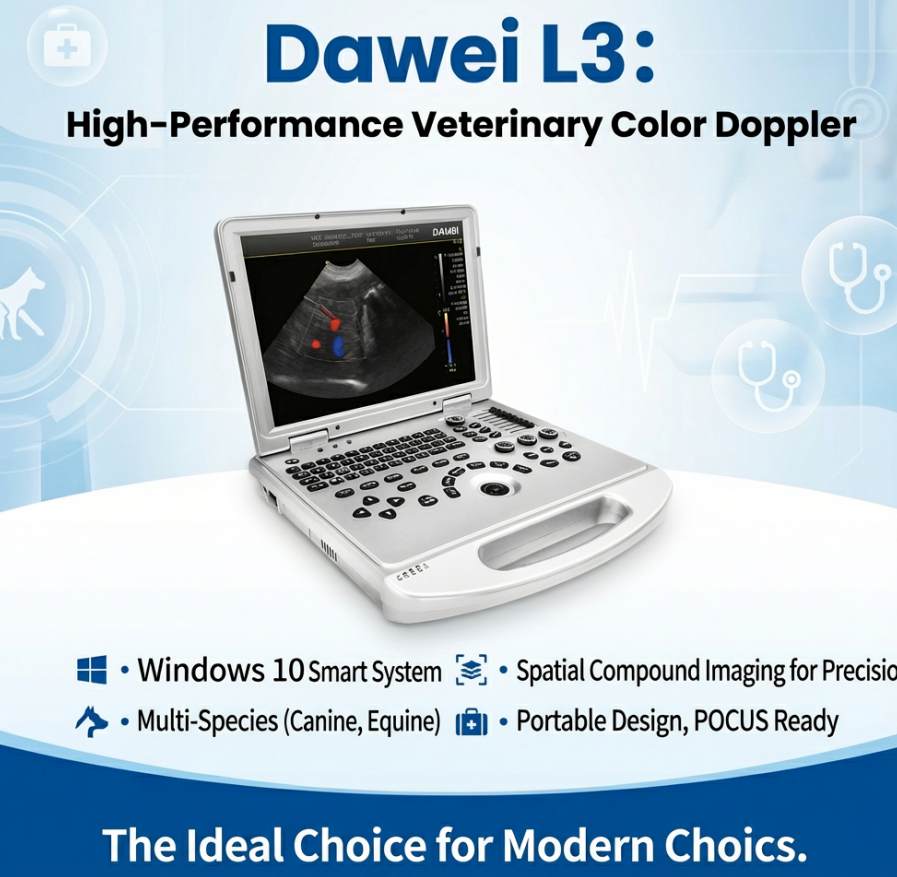
Images: Portable Devices for Dog Ultrasound
Visualizing the equipment helps in understanding its form factor and interface. Below are images of the L3-VET and related portable systems from DWVET.
The DW L3-VET features a clean, professional laptop design with a full control panel including a trackball and specialized time-gain compensation (TGC) sliders.
FAQ: Choosing and Using Portable Dog Ultrasound
Here are answers to the most common questions veterinarians ask when considering a portable ultrasound upgrade.
1. How do I choose the best portable ultrasound under 5kg for my practice?
Focus on the balance between weight and image quality. Ensure the device has a dedicated veterinary software package, sufficient battery life (2+ hours), and essential imaging modes like Color Doppler. Avoid generic human-use machines that lack specific animal presets.
2. Is a laptop-style ultrasound better than a handheld wireless probe?
For comprehensive exams, yes. Laptop units generally offer better processing power, longer battery life, and more physical controls for fine-tuning images. Handheld probes are excellent for quick “triage” scans but may lack the depth of features needed for a full diagnostic workup.
3. Which probe is most essential for a general dog practice?
The micro-convex probe is the most versatile. It fits between ribs and provides a wide field of view for abdominal organs in small to medium dogs. A linear probe is a good secondary option for superficial structures.
4. Can I export images to my clinic’s practice management software?
Yes, provided the machine supports DICOM 3.0. This allows the ultrasound to communicate directly with your PACS server. Alternatively, you can export images as JPEGs via USB.
5. How do I clean and maintain a portable ultrasound probe?
Use manufacturer-approved disinfectant wipes that do not contain alcohol or bleach, which can damage the lens. Inspect the cable regularly for cracks, especially if the device is frequently moved.
6. What training resources are available for new users?
Most reputable suppliers like DWVET offer user manuals, video tutorials, and sometimes online or in-person training sessions. The L3-VET also features built-in presets that help new users get a good image quickly.
Post time: Jan-29-2026