


Pet ultrasound examination plays a crucial role in the medical diagnosis of dogs and cats, particularly in the context of the urinary system. External manifestations of urinary system diseases often manifest as abnormal urination. However, the causes of urinary abnormalities are diverse, including tumors, stones, inflammation, and the affected areas vary, such as the bladder, urethra, and kidneys. Therefore, in the diagnosis of dogs and cats, a comprehensive examination of each part of the urinary system is essential.
1. Renal Ultrasound Detection:
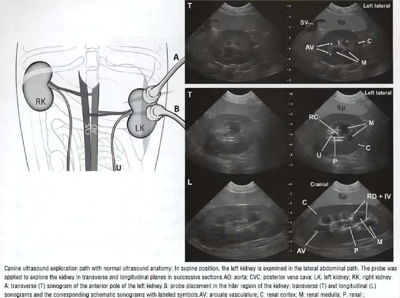
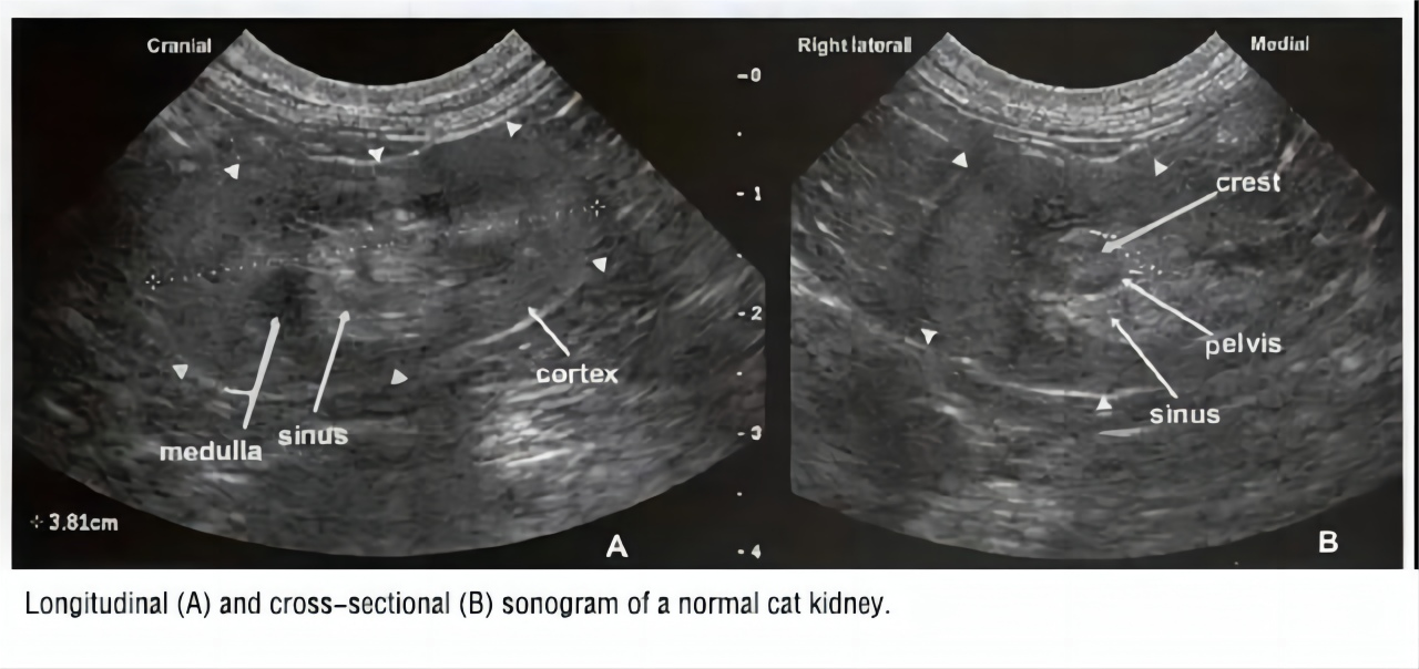
Positioning: When conducting renal detection in dogs and cats, it is essential to keep them in a comfortable and quiet supine position. Appropriate left and right lateral positioning methods can be selected.
Probe Detection Position: The probe should be placed on the animal’s lumbar back, along both sides of the spine. Typically, the optimal left and right detection areas are between the tenth and thirteenth ribs, to obtain clear images of the kidneys.
Probe and Frequency Selection: Choosing an appropriate frequency probe is crucial to ensure image clarity. For renal ultrasound, a convex or sector probe is generally selected. For small animals (<10kg) and cats, a 7.5MHz or 10.0Hz probe is used, for medium-sized dogs, a 5.0MHz probe is employed, and for large dogs, a 3.0MHz or lower frequency probe is chosen for higher resolution.
Renal Detection Slice Selection: Different sections provide a comprehensive understanding of the structure and function of the kidneys. Transverse and coronal sections are commonly used angles that present the overall appearance and vascular distribution of the kidneys.
2. Bladder and Urethral Ultrasound Examination:
Positioning: For ultrasound examination of the bladder and urethra, animals can be maintained in a supine or appropriate lateral position. Ensure that the animal is relatively relaxed to better observe the bladder and urethra.
Probe Detection Position: Place the probe on the animal’s abdomen, near the pelvic region. For the bladder, the lowest point of the abdomen is examined, and for the urethra, a step-by-step scan can be performed in the pelvic region.
Probe and Frequency Selection: In bladder and urethra examinations, choosing a convex or micro-convex probe with a frequency of 3.5 to 7.5MHz typically provides sufficient depth and clarity.
Detection Conditions: During bladder and urethra ultrasound examinations, conditions with a filled bladder can be used to more clearly display the morphology and structure of the bladder. Adjust ultrasound machine parameters such as gain and depth to obtain the best image quality.
By comparing with normal images and observing changes in echo strength shown on the sonogram, urinary system diseases in dogs and cats can be easily detected using ultrasound. These include renal enlargement, renal atrophy, renal stones, urethral stones, bladder stones, renal tumors, bladder tumors, renal pelvis pyo/hydro, bladder pyo/hydro, and more.
Pet ultrasound examination of the urinary system in dogs and cats is highly beneficial for early detection and assessment of these diseases, providing essential information for veterinarians to formulate appropriate treatment plans. However, the final diagnosis may require a comprehensive evaluation combining clinical symptoms, laboratory tests, and other imaging studies.
If your veterinary clinic requires a pet color ultrasound machine or a pet ultrasound diagnostic machine for examining and diagnosing various aspects of the pet’s urinary system, digestive system, reproductive system, etc., feel free to contact us for consultation!
Post time: Dec-26-2023