


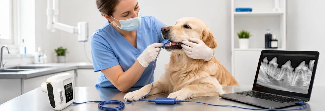
Dental problems are common in dogs and cats, but many oral diseases cannot be fully identified through a visual examination alone.
A pet’s teeth may look normal from the outside while problems such as tooth root infection, periodontal disease, or bone loss are developing below the gum line.
This is why veterinary dental radiography has become an important diagnostic tool in modern veterinary dentistry.
By using a digital dental X-ray system, veterinarians can capture detailed images of teeth, roots, and surrounding structures, helping them make more accurate treatment decisions.
What Is Veterinary Dental Radiography?
Veterinary dental radiography is a specialized imaging technique used to examine the internal structures of animal teeth and oral tissues.
Unlike a routine oral examination, dental X-rays allow veterinarians to evaluate areas that cannot be directly seen, including:
- Tooth roots
- Periodontal structures
- Jaw bone condition
- Hidden infections
- Root abnormalities
For dogs and cats, dental radiography is especially important because many dental diseases begin below the gum line.
A professional veterinary dental X-ray system provides veterinarians with clearer images and more reliable diagnostic information during oral examinations.
How Does a Veterinary Dental X-Ray System Work?
A complete dental X-ray workflow usually includes four main steps:
- Dental sensor placement
- X-ray exposure
- Digital image generation
- Diagnosis and treatment planning
Each step affects the final image quality and diagnostic accuracy.
Step 1: Dental Sensor Placement
The first step in dental radiography is positioning the intraoral sensor correctly.
The dental sensor is placed inside the animal’s mouth near the target tooth area.
Correct placement is important because it affects:
- Image clarity
- Tooth length accuracy
- Root visibility
- Diagnostic confidence
Veterinary dental sensors are designed to capture X-ray signals from inside the oral cavity and transfer the information into digital images.
During examination, veterinarians need to consider:
- Tooth position
- Sensor angle
- Animal head position
- X-ray beam direction
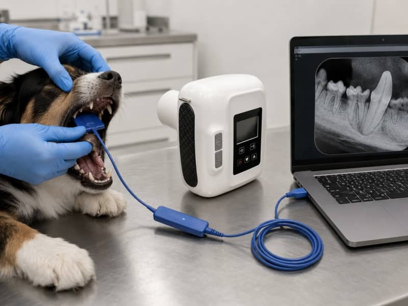
Dental sensor placement in dog dental X-ray examination
Step 2: X-Ray Exposure Process
After positioning the sensor, the X-ray generator produces a controlled beam directed toward the target tooth area.
The X-ray passes through different tissues:
- Teeth
- Bone
- Soft tissue
Different structures absorb different amounts of X-ray energy, creating differences that can be captured by the digital sensor.
A stable X-ray positioning process helps reduce image distortion and improves diagnostic quality.
Step 3: Digital Image Generation
After exposure, the digital sensor converts X-ray information into an image.
The workflow is:
Dental X-ray Generator
↓
Intraoral Sensor
↓
USB Connection
↓
Computer Imaging Software
↓
Digital Dental Image
Compared with traditional film-based imaging, digital dental X-ray provides:
- Faster image acquisition
- Easier image storage
- Simple sharing and review
- Better clinical workflow
A digital dental X-ray system helps veterinary clinics manage dental cases more efficiently by connecting image acquisition and digital diagnosis.
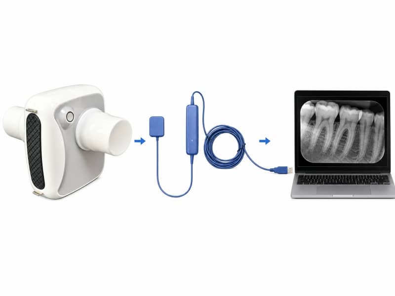
Digital dental X-ray imaging workflow
Step 4: Diagnosis and Treatment Planning
Once the dental image is generated, veterinarians can evaluate the oral condition and decide the next treatment step.
Dental X-ray images can help identify:
Tooth Root Problems
Some tooth root abnormalities cannot be detected through external examination.
Periodontal Disease
Radiographs help evaluate bone loss and changes around teeth.
Tooth Fractures
Dental imaging helps determine whether fractures extend into deeper tooth structures.
Retained Tooth Roots
After extraction procedures, X-ray imaging helps confirm whether tooth roots remain.
Why Digital Dental X-Ray Is Important for Dogs and Cats
Traditional oral examination remains an important first step, but it has limitations.
Digital dental X-ray provides additional information by showing structures hidden below the gum line.
Benefits include:
More Accurate Diagnosis
Veterinarians can evaluate tooth roots and supporting bone structures.
Faster Clinical Workflow
Digital images can be obtained, reviewed, and stored quickly.
Better Communication With Pet Owners
Clear dental images help veterinarians explain oral conditions and recommended treatments.
What Equipment Is Needed for Veterinary Dental Radiography?
A complete veterinary dental imaging workflow usually includes:
Dental X-Ray Generator
Provides controlled X-ray exposure for dental imaging.
Intraoral Dental Sensor
Captures dental images inside the animal’s mouth.
Computer and Imaging Software
Processes and stores digital dental images.
Dental Positioning Tools
Help maintain correct sensor placement and imaging angles.
For veterinary clinics performing regular dental procedures, choosing the right veterinary dental X-ray system can improve both diagnostic accuracy and workflow efficiency.
FAQ
How does veterinary dental X-ray work?
Veterinary dental X-ray works by sending X-ray beams through the tooth area and capturing the information with a digital intraoral sensor. The sensor transfers the image to computer software for diagnosis.
Where is the dental sensor placed during an X-ray?
The dental sensor is placed inside the animal’s mouth near the tooth being examined. Correct positioning helps produce clearer images.
Why are dental X-rays needed for dogs and cats?
Dental X-rays allow veterinarians to detect problems below the gum line, including root infections, periodontal disease, and hidden dental abnormalities.
Are digital dental X-rays better than traditional dental imaging?
Digital dental X-rays provide faster image acquisition, easier storage, and more efficient workflow compared with traditional film-based methods.
Conclusion
Veterinary dental radiography combines accurate sensor positioning, controlled X-ray exposure, and digital image processing to provide detailed information about animal oral health.
From sensor placement to final diagnosis, every step affects image quality and clinical decision-making.
For modern veterinary clinics, a reliable veterinary dental X-ray system helps veterinarians improve dental examinations and provide better care for dogs and cats.
Post time: Jul-27-2026