


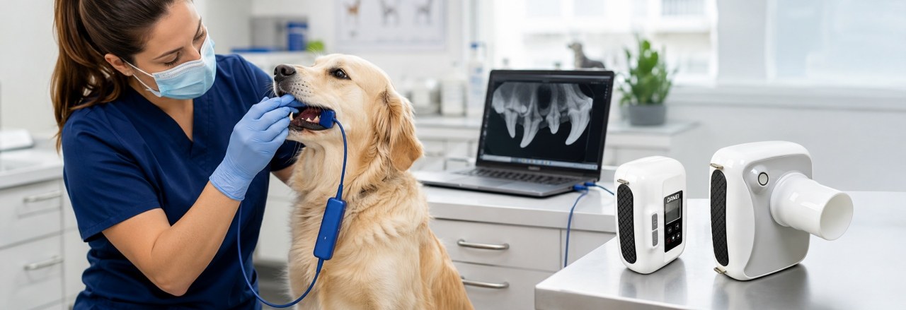
Dental disease is one of the most common health problems in dogs and cats. Although a visual oral examination can identify some dental problems, many important conditions occur below the gum line and cannot be detected without dental radiography.
Veterinary dental X-ray allows veterinarians to evaluate tooth roots, periodontal structures, jaw bones, and hidden oral problems that are not visible during a routine examination.
Understanding how to properly take dental X-rays in dogs and cats is essential for obtaining clear diagnostic images and improving treatment decisions.
Why Are Dental X-Rays Important in Veterinary Dentistry?
A normal oral examination mainly evaluates the visible part of the teeth.
However, many dental problems are hidden below the gingiva, including:
- Tooth root infection
- Periodontal disease
- Tooth fractures
- Retained tooth roots
- Jaw bone abnormalities
- Oral lesions
Dental radiography provides information about structures that cannot be evaluated through visual examination alone.
With a digital veterinary dental X-ray system, veterinarians can quickly obtain high-quality images and store examination records for future reference.
What Equipment Is Needed for Veterinary Dental X-Ray?
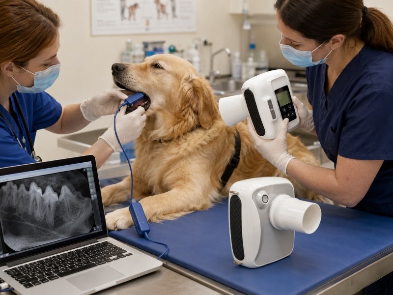
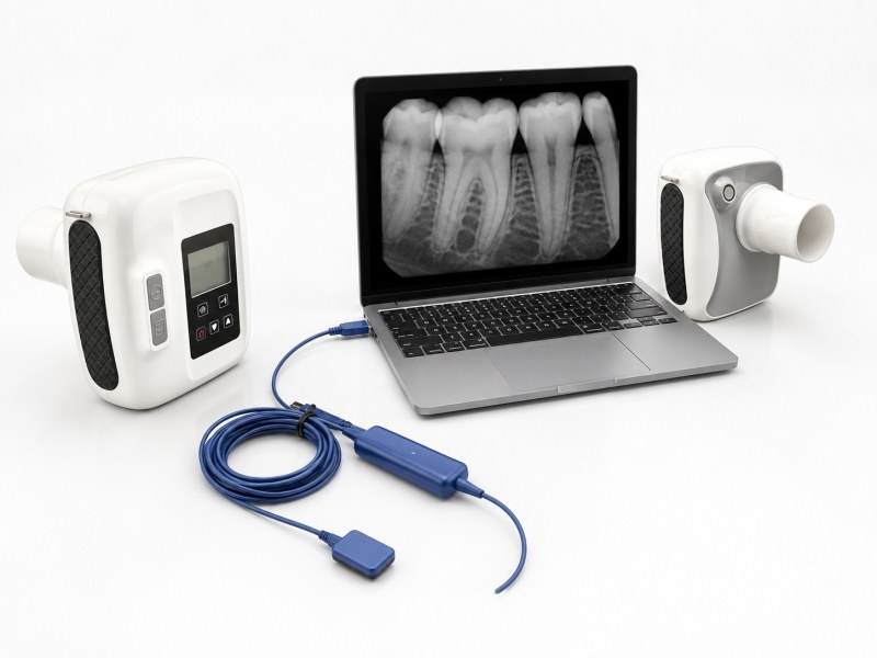
A complete veterinary dental radiography workflow usually includes:
Digital Dental X-Ray System
The X-ray generator produces radiation needed to create dental images.
Intraoral Dental Sensor
The digital sensor is placed inside the animal’s mouth to capture X-ray images.
A veterinary dental sensor should provide:
- High-resolution imaging
- Reliable positioning
- Easy cleaning and handling
Computer and Imaging Software
The sensor connects to the computer through USB to transfer dental images for viewing, storage, and diagnosis.
How Do Veterinarians Take Dental X-Rays in Dogs and Cats?
The dental X-ray procedure usually includes several steps.
1. Patient Preparation
Before dental imaging, veterinarians usually:
- Perform oral examination
- Evaluate the dental condition
- Prepare anesthesia or sedation if required
- Position the animal safely
Proper patient positioning helps reduce movement and improves image quality.
2. Positioning the Dental Sensor
Correct sensor placement is one of the most important steps in veterinary dental radiography.
The sensor should be positioned close to the target tooth area while maintaining a stable position.
Different teeth require different positioning techniques.
Common Dental X-Ray Positioning Methods
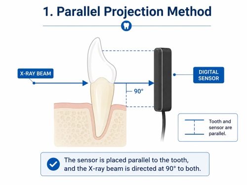
Parallel Projection Method
The parallel projection method is used when the dental sensor can be placed parallel to the target tooth.
The X-ray beam is directed perpendicular to both the tooth and the sensor.
Advantages:
- Reduced image distortion
- Better tooth length representation
- Improved diagnostic accuracy
- Areas where the sensor can be positioned stably
Bisecting Angle Projection Method
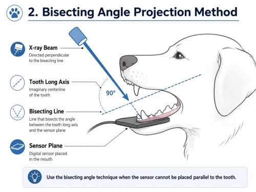
When the sensor cannot be positioned parallel to the tooth, the bisecting angle technique can be used.
The X-ray beam is directed perpendicular to an imaginary line that divides the angle between the tooth and sensor.
This method helps obtain a more accurate representation of tooth structure.
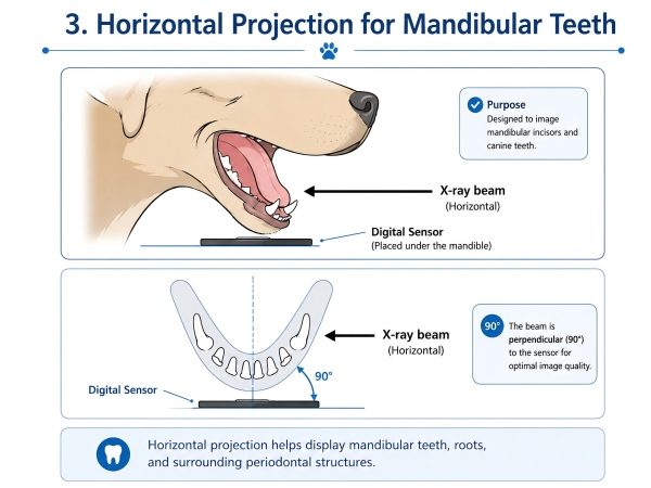
Horizontal Projection for Mandibular Teeth
Horizontal projection is commonly used for mandibular dental imaging.
Proper sensor placement and beam direction help:
- Reduce overlapping structures
- Improve visualization of tooth roots
- Evaluate periodontal structures
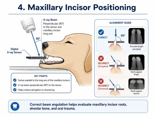
Maxillary Incisor Positioning
For maxillary incisors, stable head positioning and correct sensor placement are important.
The X-ray beam angle should be adjusted carefully to avoid:
- Tooth elongation
- Tooth shortening
- Image distortion
This allows veterinarians to evaluate:
- Incisor roots
- Surrounding bone
- Oral trauma
-
How Digital Dental X-Ray Improves Veterinary Diagnosis
Compared with traditional dental imaging methods, digital dental X-ray provides several advantages:
Faster Image Acquisition
Digital sensors allow veterinarians to obtain images quickly during dental procedures.
Better Diagnostic Visibility
High-resolution images help evaluate:
- Tooth roots
- Bone structures
- Hidden dental disease
Easy Image Management
Digital images can be:
- Stored
- Reviewed
- Shared with owners
- Used for medical record
Digital images captured by a digital dental X-ray system can be stored, reviewed, shared with pet owners, and used for medical records.
Common Dental Problems Diagnosed with Veterinary Dental X-Ray
Veterinary dental radiography helps identify:
Periodontal Disease
Dental X-rays help evaluate bone loss around teeth.
Tooth Root Problems
Radiography can reveal root abnormalities that cannot be seen externally.
Tooth Fractures
Images help determine whether fractures affect the tooth root.
Retained Tooth Roots
Dental X-ray is important after extractions to confirm complete removal.
Choosing the Right Veterinary Dental X-Ray System
When selecting a dental X-ray system for a veterinary clinic, consider:
Image Quality
Clear images are essential for accurate dental diagnosis.
Sensor Size and Handling
The sensor should fit different animal sizes and allow flexible positioning.
Workflow Efficiency
A complete system should support:
- Fast image acquisition
- Easy operation
- Digital storage
For veterinary clinics performing regular dental procedures, a professional digital dental X-ray system can significantly improve diagnostic workflow.
FAQ
Do dogs need dental X-rays before dental treatment?
Yes. Dental X-rays help veterinarians evaluate tooth roots and hidden problems before treatment.
Can dental X-rays detect problems below the gums?
Yes. Dental radiography can show tooth roots, bone structures, and diseases that cannot be seen during normal examination.
What is the difference between dental X-ray and normal veterinary X-ray?
Dental X-ray systems are specifically designed for detailed imaging of teeth and oral structures, while general veterinary X-ray systems are used for larger body areas.
How does a veterinary dental sensor work?
A digital dental sensor captures X-ray signals and transfers images to a computer through a USB connection for diagnosis and storage.
Conclusion
Dental X-rays have become an essential part of modern veterinary dentistry.
By using correct sensor positioning techniques and a reliable digital dental X-ray system, veterinarians can obtain clearer images, identify hidden dental problems, and provide better treatment for dogs and cats.
A professional veterinary dental radiography workflow helps clinics improve diagnostic confidence and deliver higher-quality oral care.
Post time: Jul-24-2026