


Why litter size matters in sheep production
Accurately distinguishing single vs. multiple fetuses helps you:
- Feed correctly (avoid underfeeding twins/triplets and overfeeding singles)
- Reduce metabolic disease risk (pregnancy toxemia, hypocalcemia)
- Plan lambing supervision (more help for multiples)
- Group ewes smarter (single-bearing vs. multiple-bearing management)
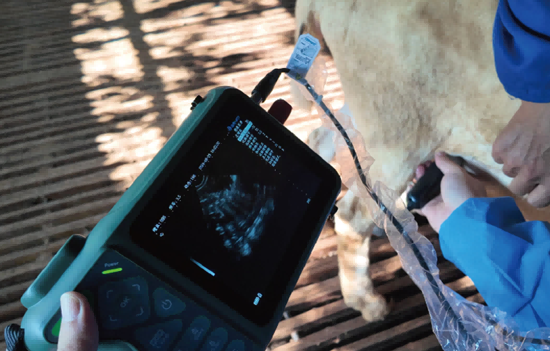
If you’re scanning on-farm and want an easy-to-carry device, you can jump to Elite handheld veterinary ultrasound and then come back to the scanning steps.
Best time window to count fetuses (and why timing matters)
Counting is easiest when the uterus is accessible and fetal structures are separated enough to recognize.
| Goal | Recommended gestation window (days) | What you can reliably see | Notes |
| Confirm pregnancy | 25–35 | Gestational sac(s), early embryo | High risk of confusing fluid pockets; be systematic |
| Count fetuses (best balance) | 40–70 | Fetal body, heartbeat, head/torso separation | Most practical on farm |
| Confirm viability / late checks | 70–120+ | Larger fetuses, placentomes | Counting becomes harder as fetuses overlap |
Practical tip: If your main goal is litter size, aim for ~45–65 days.
Anatomy + probe placement: where to scan the ewe
Most sheep pregnancy scanning is done transabdominally (through the belly).
Positioning checklist
- Restraint: standing in a crate/stand is ideal
- Hair/wool: clip or part wool if heavy; use plenty of gel
- Probe location: start just in front of the udder, slightly to the side of midline
- Direction: aim the probe cranially (toward the head) and sweep slowly
Quick scanning map (field-friendly)
- Start right inguinal area (near udder base)
- Sweep cranially along the abdominal wall
- Repeat on the left side
- Return to the side where you saw the clearest uterus and count systematically
The most reliable ultrasound signs for single vs. multiple fetuses
1) Count separate fetal bodies, not “heads you think you saw”
The most dependable rule is: a fetus = a fetal body with its own heartbeat.
What to look for:
- Distinct fetal torso outline
- Visible rib/spine line in later mid-gestation
- Heartbeat (use B-mode motion; some devices may offer M-mode)
- A single fetus may move and appear in multiple planes
- Multiples will show two (or more) heartbeats that can be found in different positions during the same scan session
2) Identify separate heartbeats in different locations
3) Use placentomes as “supporting evidence” (not proof)
Placentomes (placental attachment sites) increase with pregnancy and may be more numerous with multiples, but:
- they are not a direct fetus count
- visibility depends on gestational age and operator skill
4) Look for two amniotic sacs / membranes early on
In earlier windows (around 30–45 days), you may see:
- separate fluid sacs
- dividing membranes
This can suggest multiples, but confirm later if management decisions are high-stakes.
Step-by-step method to avoid double-counting
Double-counting is the #1 reason producers get “twins” that lamb as singles.
A simple, repeatable counting protocol
- Find the uterus and freeze your first clear fetal image
- Locate heartbeat and note position (right/left, shallow/deep)
- Sweep away and find a second fetus with a heartbeat in a different location
- If unsure, re-scan the same ewe from the other side
- Record as: Single / Twin / Triplet+ / Uncertain
Recording template (use in a notebook or app)
| Ewe ID | Days bred (est.) | Result | Confidence | Notes |
| 1023 | 55 | Twin | High | Two separate heartbeats; right + left |
Visual cues by gestational stage (what you’ll typically see)
| Gestation stage | Single pregnancy tends to look like | Multiple pregnancy tends to look like |
| 25–35 days | One sac, one embryo | Two sacs or two embryos, may overlap |
| 40–70 days | One fetus fills most of view in one sweep | Two fetal bodies appear with careful sweeps; heartbeats in distinct areas |
| 70–120+ days | Large fetus, overlap can confuse | Fetuses overlap; counting becomes less accurate |
Common mistakes (and how to fix them)
- Mistake: confusing one fetus in two planes as twins
Fix: only count a second fetus if you can confirm a separate heartbeat in a different location. - Mistake: scanning too late (fetuses overlap)
Fix: schedule scanning earlier for litter size (often 45–65 days). - Mistake: too little gel / poor contact through wool
Fix: clip a small window or part wool; use more gel. - Mistake: moving too fast
Fix: slow sweeps; pause when you see the uterus and let motion reveal the heartbeat. - Mistake: relying on placentomes alone
Fix: treat placentomes as supportive; count fetuses by bodies + heartbeat.
Equipment checklist for sheep pregnancy scanning
| Item | Why it matters | Field tip |
| Ultrasound unit | Image clarity affects counting accuracy | A lightweight handheld device reduces fatigue |
| Sheep-appropriate probe | Matches scanning depth and field of view | Many sheep scans use a mechanical sector/convex type |
| Gel | Prevents air gaps | Carry extra; cold weather uses more gel |
| Restraint setup | Safety + scan speed | A simple standing crate works well |
| Record sheet/app | Prevents mix-ups | Record confidence level (High/Medium/Low) |
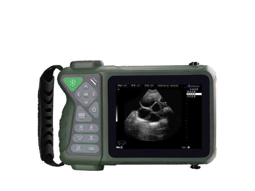
Key Elite specs (from the technical parameters)
| Specification | Elite value |
| Display | 5.6-inch LED |
| Scan method | 3.5M mechanical sector scan |
| Languages | Chinese / English UI |
| Display modes | B, B+B, 4B |
| Body position markers | ≥17 |
| Frequency options | 2.0 / 2.5 / 3.5 / 4.0 / 5.0 MHz |
| Cine loop | ≥256 frames (continuous or frame-by-frame) |
| Storage | Built-in 8GB + USB flash drive support |
| Gray scale | 256 levels |
| Dynamic range | 0–135 dB |
| Depth | 126–307 mm |
| Battery | 2600 mAh removable rechargeable Li-ion; ~3 hours runtime |
| Size | 240 × 120 × 45 mm |
| Weight | ~900 g |
How Elite fits the sheep litter-size workflow
- Mid-gestation counting (40–70 days): the depth range and multiple frequencies can help optimize clarity.
- Fast farm use: cine loop (movie playback) helps you re-check heartbeats without re-scanning.
- Training new staff: ≥17 body markers and simple B-mode options support repeatable positioning.
Internal linking tip for your site: link earlier mentions like “handheld veterinary ultrasound for sheep” to this S0 section.
FAQ: Sheep ultrasound fetal counting
1) What is the best day to ultrasound ewes for twins?
For most farms, around 45–65 days is the best window to differentiate single vs. multiple fetuses with good accuracy.
2) Why did my scan show twins but she lambed a single?
Most often it’s double-counting the same fetus in different planes. Use the rule: a second fetus must have a heartbeat in a different location.
3) Can I count triplets reliably with ultrasound?
Sometimes, but accuracy drops as litter size increases and fetuses overlap. Many operators record “Triplet+” when three distinct fetal bodies/heartbeats are confirmed.
4) Do placentomes tell you how many lambs there are?
Not reliably. Placentomes indicate pregnancy and placental development, but they are not a direct fetus count.
5) Standing or lying—what’s better for scanning sheep?
Standing restraint is common for farm scanning because it’s fast and repeatable. What matters most is consistent probe placement and good contact.
Post time: Mar-19-2026