


Veterinary DR Selection Guide: Deep Dive into Detector Size, Pixel Pitch, and Clinical Image Quality
In the digital upgrade of veterinary imaging departments, the Flat Panel Detector (FPD)—the core component of Digital Radiography (DR) systems—directly determines imaging quality and workflow efficiency. For many veterinary clinic owners, facing the complex array of specifications on the market—such as “14×17 vs 17×17 inches,” “100µm vs 140µm pixel pitch,” and professional terms like “MTF/DQE”—makes it difficult to choose the system that best matches clinical needs. This article strips away marketing jargon to deeply analyze the real impact of size and resolution on animal diagnostic imaging from both physical and clinical perspectives.
In short, Detector Size determines “how large an animal you can image” and “whether you need to frequently adjust the equipment,” directly affecting positioning efficiency for large dogs. Meanwhile, Resolution is not solely determined by pixel count; it is a comprehensive metric composed of Pixel Pitch, Modulation Transfer Function (MTF), and Detective Quantum Efficiency (DQE). For most veterinary clinical tasks, blindly pursuing extremely small pixels is unnecessary; balancing dose efficiency with clinical sharpness is the key.
Executive Summary
- Detector Size & Positioning: Detector size drives field of view and positioning efficiency. A 17×17 inch panel provides full coverage, significantly reducing rotation requirements during thoracic and abdominal exams for large dogs.
- Resolution Truth: Clinical sharpness is limited by the Nyquist frequency upper bound but depends more on MTF (contrast transfer) and DQE (dose efficiency). Resolution is a function of pixel pitch, Nyquist, MTF, and DQE.
- Selection Advice: For typical veterinary DR tasks, a balanced pixel pitch (e.g., 140µm) combined with a high DQE (CsI) scintillator and effective dose management matters most for general practice (orthopedics, soft tissue).
1.Detector Size: Field of View, Positioning, and Coverage
In veterinary DR systems, the two most common physical detector sizes are 14×17 inches (approx. 35×43 cm) and 17×17 inches (approx. 43×43 cm). This seemingly minor 3-inch difference has a massive impact on actual veterinary clinical positioning workflow.
For cats, small dogs, or exotics, a 14×17 inch panel is more than sufficient. However, when facing the thoracicabdominal imaging of medium-to-large breeds like Golden Retrievers, German Shepherds, or Labradors, a 14×17 inch panel often fails to cover the entire target area in a single shot. If using a 14×17 panel, technicians may need to place it transversely for the chest and then rotate it longitudinally for the abdomen, or perform multiple exposure stitching. This not only increases radiation dose but also leads to motion artifacts due to animal movement caused by frequent panel adjustments. In contrast, a 17×17 inch large field-of-view panel can accommodate the chest and abdominal structures of most adult large dogs in one go, eliminating the need to rotate the detector and greatly improving throughput in emergency and high-volume clinics.
Table 1: Veterinary DR Detector Size Comparison and Clinical Applicability
| Size (Inches) | Effective FOV (cm) | Typical Pixel Pitch | Nyquist (lp/mm) | Positioning Efficiency | Common Use Cases |
| 14 × 17 | 35 × 43 | 140 – 150 µm | ~3.3 – 3.5 | Medium (Rotation required for large animals) | Mobile/Portable, Cat/Small Dog Specialists, Retrofits |
| 17 × 17 | 43 × 43 | 100 – 140 µm | ~3.5 – 5.0 | High (Omni-directional, no rotation) | Fixed DR Tables, Large Breed General Practice, High-Volume Hospitals |
2.Resolution Mechanics: MTF, DQE, and Pixel Pitch
Many buyers mistakenly believe that “smaller pixels mean clearer images,” which is physically one-sided. According to the Nyquist sampling theorem, Pixel Pitch (p) does determine the system’s limiting spatial resolution, calculated as Nyquist Frequency = 1 / (2 × p). For example, a 100µm pixel has a limiting resolution of 5 lp/mm, while 140µm is 3.57 lp/mm. However, in actual clinical imaging, we rarely reach this theoretical upper limit.
Real image sharpness is more dependent on the system’s Modulation Transfer Function (MTF) and Detective Quantum Efficiency (DQE). As noted in medical physics research, if pixels are too small, the number of photons received by a single pixel decreases, leading to a drop in Signal-to-Noise Ratio (SNR). To maintain image quality, radiation dose must be increased, which contradicts the ALARA (As Low As Reasonably Achievable) principle. Furthermore, an Agfa white paper study indicates that within the 76µm to 150µm range, pixel size has minimal impact on “perceived clinical image quality”; instead, dose levels and low-frequency DQE are the dominant factors. For veterinarians, selecting a Cesium Iodide (CsI) scintillator with high DQE is more clinically valuable than simply pursuing extremely small pixels.
Core Definitions:
Smaller pixel pitch (µm) raises the Nyquist limit (lp/mm), but clinical sharpness also depends on MTF and DQE.
- Pixel Pitch: Distance between the centers of adjacent pixels. Smaller pitch means higher theoretical resolution.
- Nyquist Frequency: The highest frequency a digital system can resolve, measured in line pairs per millimeter (lp/mm).
Quality Indicators:
- MTF (Modulation Transfer Function): The system’s ability to preserve contrast at various details, reflecting “sharpness”.
- DQE (Detective Quantum Efficiency): The efficiency of converting X-rays into image signals, reflecting “dose efficiency”.
3.Device Example: DAWEI RV-32B Specs and Clinical Meaning
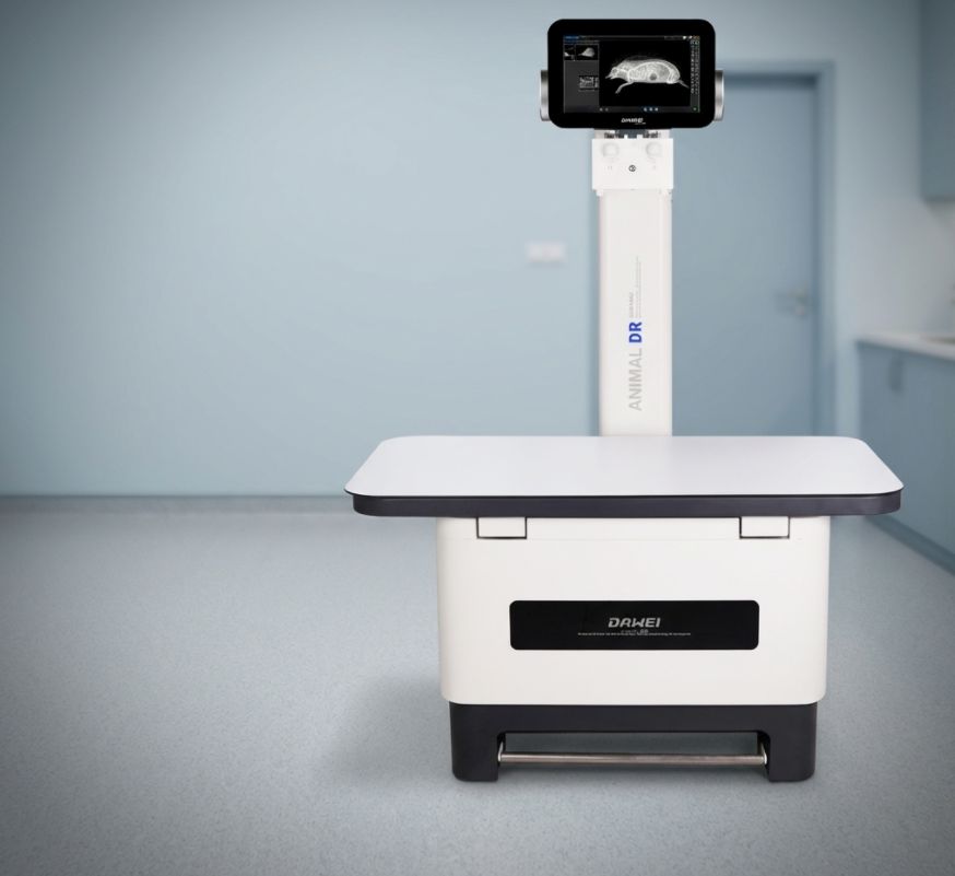
To illustrate how these theories apply to actual products, we look at the DAWEI RV-32B Veterinary Digital X-ray System. This device employs a large-format, high-sensitivity flat panel detector, with engineering specifications that exemplify the “Large FOV” and “Balanced Resolution” philosophy.
Table 2: DAWEI RV-32B Core Imaging Specifications
|
Parameter |
Specification Value | Clinical Meaning |
| Tabletop Size | 1400 × 720 mm | Ample working area for large breeds |
| Output / Tube | 32 kW / 10-400 mA | High penetration for dense bone or obesity |
| Imaging Area | 430 × 430 mm (17×17″) | Covers large dog thorax/abdomen; no rotation needed |
| Scanning Matrix | 3072 × 3072 pixels | ~9.4 Megapixels of high definition detail |
| Pixel Size | 140 µm | Balanced resolution (Nyquist ~3.57 lp/mm) & low noise |
| Scintillator | CsI (Cesium Iodide) |
High DQE reduces required dose, improving sharpness |
Spec Analysis: The RV-32B selects a 140 µm pixel size, which is a time-tested “Golden Balance” in veterinary radiology. While pixels smaller than 100µm theoretically offer finer trabecular bone detail, for routine imaging (e.g., abdominal organ layers, fracture assessment), 140µm combined with a CsI scintillator provides excellent MTF characteristics. This ensures sharp edges while avoiding the high noise issues associated with extremely small pixels. The 3072×3072 matrix paired with a 17×17 inch FOV makes it a true “All-Rounder” for veterinary practice, capable of capturing minute renal calculi in cats as well as full hip dysplasia (HD) evaluations in large dogs.
4. Veterinary Clinic Environment
The image below demonstrates the deployment of the DAWEI RV-32B in a standard veterinary exam room. The workstation. This integrated design minimizes cable interference, facilitating rapid animal restraint and positioning. 1400x720mm floating tabletop combined with the integrated 17×17 inch detector creates an efficien.
5.Selection Strategy: Pixel vs. FOV vs. Dose
Veterinary imaging departments should formulate strategies based on their specific caseload. Blindly pursuing a single specification often leads to wasted budget or operational inconvenience.
- When to prioritize Small Pixels (≤100–125 µm)? When your clinic predominantly treats exotic pets (hamsters, birds, lizards) or focuses on dentistry and distal micro-fractures. These tiny anatomical structures demand high-frequency spatial resolution, making the trade-off of slightly lower dose efficiency for detail worthwhile.
- When to prioritize Large FOV (17×17 inch)? For General Veterinary Hospitals, Emergency Centers, or Orthopedics. Full spine or thorax-abdomen studies of medium-to-large dogs are daily high-frequency items. A 17×17 inch panel significantly speeds up workflow and reduces retakes.
- Dose Management Baseline: Regardless of size, CsI (Cesium Iodide) scintillators should be standard. Unlike GoS (Gadolinium Oxysulfide), CsI has a needle-like crystal structure that drastically reduces light scatter and improves DQE, which is crucial for protecting veterinary staff who often need to perform manual restraint.
- Geometry & Standards: Remember that image quality also depends on geometry. Using a grid for thick body parts (>10cm) and maintaining correct Source-Image Distance (SID) is vital. As noted in IEC 62220-1 standards, MTF/DQE measurements assume specific beam qualities; ensure your clinical setup mimics standard geometry for optimal results.
6.Applications & Compliance
High-performance veterinary DR systems like the RV-32B are widely applicable in the following clinical scenarios:
- Small Animal Thorax/Abdomen: Rapid capture of cardiopulmonary details; assessment of pleural effusion, tumors, and foreign bodies.
-Orthopedics: Pre-operative planning (TPLO, fracture internal fixation) and post-operative healing assessment.
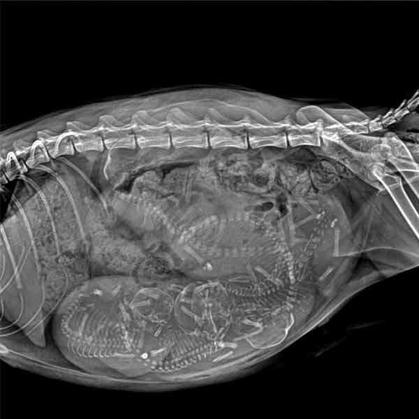
-Gastrointestinal Studies: Barium contrast studies to evaluate peristalsis and obstruction. -Reproduction & Exotics: Gestational fetal counting; dystocia risk assessment; dental and exotic pet imaging.
Safety & Compliance Reminder: No matter how high the resolution, image quality relies on standard operation. Always adhere to Radiation Protection principles (Time, Distance, Shielding/ALARA). Perform regular Gain/Offset Calibration and Quality Assurance/Quality Control (QA/QC) on the DR system to ensure consistent output of diagnostic-quality images.
Post time: Jan-08-2026